Adductor Magnus
Description
-
![Schematic image illustrating the adductor minimus muscle and surrounding landmarks. Note the arrangement of the arteries around the borders of this muscle [1]](/w/images/thumb/8/81/Adductor_magnus_vascular.jpeg/77px-Adductor_magnus_vascular.jpeg) Schematic image illustrating the adductor minimus muscle and surrounding landmarks. Note the arrangement of the arteries around the borders of this muscle [1]
Schematic image illustrating the adductor minimus muscle and surrounding landmarks. Note the arrangement of the arteries around the borders of this muscle [1] -
![The posteromedial corner of the knee. The adductor magnus is marked AMT[2]](/w/images/thumb/6/62/Adductor_magnus_tendon.jpeg/120px-Adductor_magnus_tendon.jpeg) The posteromedial corner of the knee. The adductor magnus is marked AMT[2]
The posteromedial corner of the knee. The adductor magnus is marked AMT[2] -
-
-
-
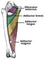
![Observed attachments of psoas, iliacus, pectineus, adductor brevis and adductor magnus (a, posteromedial view; b, posterior view)[3]](/w/images/thumb/c/c1/Muscle_attachments_medial_thigh.jpeg/120px-Muscle_attachments_medial_thigh.jpeg) Observed attachments of psoas, iliacus, pectineus, adductor brevis and adductor magnus (a, posteromedial view; b, posterior view)[3]
Observed attachments of psoas, iliacus, pectineus, adductor brevis and adductor magnus (a, posteromedial view; b, posterior view)[3]
![Schematic image illustrating the adductor minimus muscle and surrounding landmarks. Note the arrangement of the arteries around the borders of this muscle [1]](/File:Adductor_magnus_vascular.jpeg)
![The posteromedial corner of the knee. The adductor magnus is marked AMT[2]](/File:Adductor_magnus_tendon.jpeg)


![Observed attachments of psoas, iliacus, pectineus, adductor brevis and adductor magnus (a, posteromedial view; b, posterior view)[3]](/File:Muscle_attachments_medial_thigh.jpeg)


Introduction
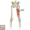
- Large triangular muscle located in the medial compartment of the thigh
- Primarily an adductor muscle, also contributes to some flexion and extension
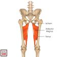
Origin
- Superior segment: ramus of the pubis
- Inferior segment: ramus of the ischium [6]
Insertion
- Superior segment: medial gluteal tuberosity (medial to the Gluteus Maximus)
- Inferior segment: linea aspera and proximal medial supracondylar line of the femur [6] [7]
Actions
- Adductor portion
- Adduction of the thigh
- Flexion of the thigh
- Hamstring Portion
- Adduction of the thigh
- Extension of the thigh
- Both portions work together during the gait cycle and control the pelvis
Vascular
- Primary source: Perforating branches of the profunda femoris artery
- Superior portion: medial femoral circumflex
- Inferior portion: popliteal and genicular arteries
- Other arteries: Obturator artery
Nerve
- Posterior branch of the obturator nerve
- Tibial portion of the sciatic nerve [7]
- Adductor magnus forms part of the posterior border of Adductor Canal along with adductor longus
Clinical Significance
Pathology
Procedure
See Also
References
- ↑ Shane Tubbs, R., et al. "The adductor minimus muscle revisited." Surgical and radiologic anatomy 33.5 (2011): 429-432.
- ↑ Cinque, Mark E., et al. "Posteromedial corner knee injuries: diagnosis, management, and outcomes: a critical analysis review." JBJS reviews 5.11 (2017): e4.
- ↑ Sedlmayr, Jayc C., et al. "Revision of hip flexor anatomy and function in modern humans, and implications for the evolution of hominin bipedalism." The Anatomical Record 305.5 (2022): 1147-1167.
- ↑ Broski, Stephen M., et al. "The adductor magnus “mini-hamstring”: MRI appearance and potential pitfalls." Skeletal Radiology 45 (2016): 213-219.
- ↑ Lungu, Eugen, Johan Michaud, and Nathalie J. Bureau. "US assessment of sports-related hip injuries." Radiographics 38.3 (2018): 867-889.
- ↑ 6.0 6.1 Jeno SH, Schindler GS. Anatomy, Bony Pelvis and Lower Limb, Thigh Adductor Magnus Muscles. [Updated 2018 Dec 16]. In: StatPearls [Internet]. Treasure Island (FL): StatPearls Publishing; 2020 Jan-.
- ↑ 7.0 7.1 Takizawa M, Suzuki D, Ito H, Fujimiya M, Uchiyama E. The adductor part of the adductor magnus is innervated by both obturator and sciatic nerves. Clin Anat. 2014 Jul;27(5):778-82.
Created by:
Connor Farrell on 10 February 2020 19:01:07
Authors:
Last edited:
17 June 2026 18:43:58
Category: