Calcaneal Apophysitis
Other Names
- Sever's Disease
- Traction Apophysitis Injury of the Ankle
- Calcaneal Apophyseal Avulsion Fracture
Background
- This page covers apophysitis of the calcaneus, a cause of heel pain seen in kids often referred to as Sever's Disease
History
- First reported in 1912 by James Warren Severe, an orthopedic specialist[1]
- In 1926, Lewin first postulated a cause[2]
Epidemiology
- The most common cause of heel pain in pediatrics
- Primarily seen between the ages of 8 and 15
- Median age: males (12 years), females (11 years) (need citation)
- Has been observed in children as young as 6[3]
- Reported incidence of 3.7 per 1,000 patients[4]
- Up to 60% of cases are bilateral (need citation)
- Reported to account for between 2% and 16% of heel pain in children presenting to sports clinics[5]
Introduction

General
- Self limiting condition which occurs due to repetitive stress at the calcaneal apophysis
- Often seen following a growth spurt in conjunction with increased sport participation
- The diagnosis is primarily clinical
Etiology
- Repetitive microtrauma
- Movement of the apophysis relative to the diaphysis causes trauma to the apophyseal tissues with compression or impact forces
- Approximately 60% of the weight-bearing load occurs in the rear foot when standing
- Sports with running and jumping report more pain (need citation)
- Growth
- Apophysis is the weakest point in the muscle-tendon-bone-attachment
- Bone growth exceeds the ability of the muscle-tendon unit to stretch sufficiently to maintain previous flexibility
- This leads to increased tension across the unossified or incompletely ossified apophysis
- Avulsion fracture
- Rarely, trauma may lead to a full avulsion fracture
Anatomy of Calcaneus
- Apophysis is the posterior aspect of the calcaneus, where the Achilles tendon inserts.
- Growth plate does not close until at least 14 years of age.
Risk Factors
- Systemic
- Sports
- Soccer
- Track and Field
- Basketball
- Cross-country
- Gymnastics
- Training/ Conditioning
- High levels of physical activity
- Heel cord tightness
- Weak ankle dorsiflexion
- Poorly cushioned or worn-out athletic shoes
- Running on hard surfaces
- Biomechanical[8]
- Genu varum
- Forefoot varus
- Pes cavus
- Pes planus
Differential Diagnosis
Differential Diagnosis Ankle Pain
- Fractures & Dislocations
- Muscle and Tendon Injuries
- Ligament Injuries
- Bursopathies
- Nerve Injuries
- Arthropathies
- Pediatrics
- Fifth Metatarsal Apophysitis (Iselin's Disease)
- Calcaneal Apophysitis (Sever's Disease)
- Triplane Fracture
- Other
Differential Diagnosis Foot Pain
- Fractures & Osseous Disease
- Traumatic/ Acute
- Stress Fractures
- Other Osseous
- Dislocations & Subluxations
- Muscle and Tendon Injuries
- Ligament Injuries
- Plantar Fasciopathy (Plantar Fasciitis)
- Turf Toe
- Plantar Plate Tear
- Spring Ligament Injury
- Neuropathies
- Mortons Neuroma
- Tarsal Tunnel Syndrome
- Joggers Foot (Medial Plantar Nerve)
- Baxters Neuropathy (Lateral Plantar Nerve)
- Arthropathies
- Hallux Rigidus (1st MTPJ OA)
- Gout
- Toenail
- Pediatrics
- Fifth Metatarsal Apophysitis (Iselin's Disease)
- Calcaneal Apophysitis (Sever's Disease)
- Freibergs Disease (Avascular Necrosis of the Metatarsal Head)
- Kohlers Disease (Avascular Necrosis of the Navicular)
Clinical Features

History
- Age is typically 8 to 15
- No specific injury
- Complain of heel pain, especially after athletic activities
- In severe cases, pain may occur at rest
- Patient may point more towards Achilles than to calcaneus
- May report a limp walking on toes
- Trouble running, jumping
- Pain is often bilateral
Physical Exam: Physical Exam Foot
- Erythema, edema are typically absent
- Tenderness with palpation or compression of medial and lateral heel
- Dorsiflexion is often limited an dpainful
Special Tests
- Calcaneal Squeeze Test: Pain is reproduced with compression of the posterior calcaneus
- Severs Sign: Aggravated by standing on tiptoes
Evaluation

Radiographs
- Standard Radiographs Foot
- Alternatively can be seen on Standard Radiographs Ankle
- May appear normal, not always diagnostic
- The diagnosis is primarily clinical, imaging is not required to confirm diagnosis
- Findings
- Increased density
- Fragmentation of the calcaneal apophysis
Classification
- Not applicable
Management

Nonoperative
- Indications
- First line in all cases
- Treatment Guidelines
- There are no clear, evidence based treatment guidelines
- Further evaluation of treatment methods is needed[12]
- Activity modification
- Must discontinue sport or offending recreational activity
- Ice therapy
- NSAIDS
- Physical Therapy
- Stretching of posterior chain and especially calf muscle and achilles tendon
- Immobilization
- May be indicated for more severer cases
- Place in Tall Walking Boot or Short Leg Cast for 2-4 weeks
- Kinesiology Taping
- Tape around the arch, heel may reduce pain
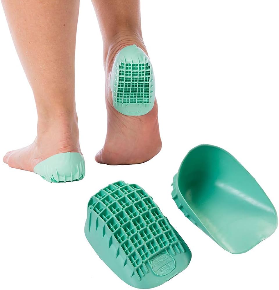
- Heel Cups
- Help absorb impact during running, jumping
- Decrease microtrauma during normal activities of daily living
- Perhamre et al found heel cups reduced pain to nearly 0/10 after about 4 weeks[13]
Operative
- Indications
- Calcaneal apophyseal avulsion fracture
- Technique
- Needs to be updated
Rehab and Return to Play
Rehabilitation
- Emphasis on stretching posterior chain, especially calf muscles
Return to Play/ Work
- Needs to be updated
Prognosis and Complications
Prognosis
- Self limited condition
Complications
- Reoccurrence of pain
- Calcaneal apophyseal avulsion fracture
- Few case reports in the literature[14]
See Also
Internal
- Pediatric Fractures (Main)
- Apophyseal And Epiphyseal Injuries (Main)
- Foot Pain (Main)
- Physical Exam Foot
- Foot Anatomy (Main)
External
- Sports Medicine Review Foot Pain: https://www.sportsmedreview.com/by-joint/foot/
References
- ↑ Sever JW: Apophysitis of the os calcis. NY Med J. 1912, 95: 1025-
- ↑ Lewin P: Apophysitis of the os calcis. Surg Gynecol Obstet. 1926, 41: 578-
- ↑ Volpon J, de Carvalho Filho G: Calcaneal apophysitis: a quantitative radiographic evaluation of the secondary ossification center. Arch Orthop Trauma Surg. 2002, 122: 338-341.
- ↑ Wiegerinck, J. I., Yntema, C., Brouwer, H. J., & Struijs, P. A. (2014). Incidence of calcaneal apophysitis in the general population. European Journal of Pediatrics, 173(5), 677–679.
- ↑ Micheli LJ, Fehlandt AF: Overuse injuries to tendons and apophyses in children and adolescents. Clin Sport Med. 1992, 11: 713-726.
- ↑ H.Chang, S.-S.Kwon, and K.-W.Minn, “Lateral calcaneal artery as a recipient pedicle for microsurgical foot reconstruction,” J. Plast. Reconstr. aesthetic Surg., vol. 63, pp. 1860–1864, 2010.
- ↑ James, A. M., Williams, C. M., Luscombe, M., Hunter, R., & Haines, T. P. (2015). Factors associated with pain severity in children with calcaneal apophysitis (Sever’s disease). The Journal of Pediatrics, 167(2), 455–459.
- ↑ McSweeney SC, Reed L, Wearing S. Foot Mobility Magnitude and Stiffness in Children With and Without Calcaneal Apophysitis. Foot Ankle Int. 2018 May;39(5):585-590.
- ↑ B. Hosgoren, A. Koktener, and G. Dilmen, “Ultrasonography of the calcaneus in Sever’s disease,” Indian Pediatr., vol. 42, p. 801, 2005.
- ↑ Case courtesy of Domenico Nicoletti, Radiopaedia.org, rID: 165100
- ↑ M.P.McHugh and C.H.Cosgrave, “To stretch or not to stretch: the role of stretching in injury prevention and performance,” Scand. J. Med. Sci. Sports, vol. 20, pp. 169–181, 2010.
- ↑ Leeb H, Stickel E: Literature review of sever’s disease: radiographic diagnosis and treatment. Podiatric Medical Review. 2012, 20: 4-9.
- ↑ Perhamre, S., et al. "A heel cup improves the function of the heel pad in Sever's injury: effects on heel pad thickness, peak pressure and pain." Scandinavian journal of medicine & science in sports 22.4 (2012): 516-522.
- ↑ Lee KT, Young KW, Park YU, Park SY, Kim KC: Neglected sever’s disease as a cause of calcaneal apophyseal avulsion fracture: case report. Foot Ankle Int. 2010, 31: 725-728. 10.3113/FAI.2010.0725.
Created by:
John Kiel on 9 March 2022 16:09:10
Authors:
Last edited:
5 August 2024 20:32:54
Categories: