Hallux Rigidus
Other Names
- Degenerative joint disease of the first metatarsophalangeal joint
- Arthritis of the first metatarsophalangeal joint
- Osteoarthritis of the first toe
- Arthritis of the first MTP
Background
- This page refers to Hallux Rigidus, a degenerative joint condition of the first Metatarsophalangeal Joint (MTPJ)
History
Epidemiology
- Most common form of arthritis of the foot[1]
- Nearly 10% of adults have symptomatic hallux rigidus[2]
- Radiographic evidence is present in 20% to 48% of adults older than 40 years
Pathophysiology
- General
- Degenerative joint disease of the 1st MTP characterized by pain, stiffness
Etiology
- General
- Primarily considered an idiopathic disease with multiple risk factors
- Underlying cause is typically multifactorial
- History of trauma
- Biomechanical factors
- Metarsus primus elevatus
- Hallux valgus
- First ray hypermobility
- Metatarsus adductus
- Non-contributatory[6]
- Achilles contracture
- Shoe wear
- Elevated metatarsal head
Pathoanatomy
- 1st Metatarsophalangeal Joint (MTPJ)
- Articulation of the first metatarsal and base of the proximal phalanx, sesamoids
- Stabilized by the joint capsule, medial and collateral ligaments, crossing musculotendinous units
Risk Factors
- Demographic
- Family History
- 2/3 of patients have a positive family history[7]
- 95% of patients with a family history had bilateral symptoms
- Women
- Family History
- Sports
- Soccer
- Runners
- Ballet
Differential Diagnosis
- Fractures & Osseous Disease
- Traumatic/ Acute
- Stress Fractures
- Other Osseous
- Dislocations & Subluxations
- Muscle and Tendon Injuries
- Ligament Injuries
- Plantar Fasciopathy (Plantar Fasciitis)
- Turf Toe
- Plantar Plate Tear
- Spring Ligament Injury
- Neuropathies
- Mortons Neuroma
- Tarsal Tunnel Syndrome
- Joggers Foot (Medial Plantar Nerve)
- Baxters Neuropathy (Lateral Plantar Nerve)
- Arthropathies
- Hallux Rigidus (1st MTPJ OA)
- Gout
- Toenail
- Pediatrics
- Fifth Metatarsal Apophysitis (Iselin's Disease)
- Calcaneal Apophysitis (Sever's Disease)
- Freibergs Disease (Avascular Necrosis of the Metatarsal Head)
- Kohlers Disease (Avascular Necrosis of the Navicular)
Clinical Features

- History
- Pain at the first metatarsophalangeal joint, especially while walking or with push off
- Pain is typically worse dorsally
- Swelling, dorsal osteophytes and soft tissue prominence
- Decreased range of motion, stiffness
- Pain while wearing tight shoes
- Pain after standing for prolonged periods
- When walking, symptoms most severe at terminal heel-rise just before toe-off
- Pain after loading 1st MTPJ such as tip-toeing, running, stairs, push ups[9]
- Antalgic gait or limp is often present
- Lateral foot pain may develop due to altered gait and walking on lateral foot
- Neuropathic pain from compression of the dorsomedial branch of the superficial peroneal nerve[10]
- Chronically, the joint may ankylose naturally and eventually become painless
- Physical Exam: Physical Exam Foot
- Swollen, inflamed first MTPJ
- Tender osteophytes on dorsal surface
- Limited range of motion
- Pain in dorsiflexion (due to dorsal osteophyte impingement)[11]
- Pain in plantarflexion (stretching of the dorsal capsule over the dorsal osteophyte)
- Decreased push off strength
- Compare to unaffected foot if symptoms unilateral
- Special Tests
- MTPJ Grind Test
- Tinels Test may indicate compression of the dorsomedial branch of the superficial peroneal nerve
Evaluation
Radiographs
- Standard Radiographs Foot
- Standard weight bearing 3 views
- Findings on lateral view
- Dorsal osteophytes
- Joint space narrowing
- Findings on AP view
- Subchondral sclerosis
- Subchondral cysts
- Flattening of the metatarsal head
- Joint space narrowing
Classification
Coughlin and Shurnas Classification
| Grade | Dorsiflexion | Radiographic findings | Clinical findings |
|---|---|---|---|
| 0 | 40-60° and/or 10-20% compared to other side | Normal | No pain; only stiffness and loss of motion on examination |
| 1 | 30-40° and/or 20-50% loss compared to other side | Dorsal osteophyte is main finding, minimal joint-space narrowing, minimal peri-articular sclerosis, minimal flattening of metatarsal head | Mild or occasional pain and stiffness, pain at extremes of dorsiflexion and/or plantar flexion on examination |
| 2 | 10-30° and/or 50-75% loss compared to other side | Dorsal, lateral and possible medial osteophytes giving flattened appearance to metatarsal head, no more than of dorsal joint space involved on lateral radiograph, mild-to-moderate joint space narrowing and sclerosis, sesamoids not usually involved | Moderate to severe pain and stiffness that may be constant; pain occurs just before maximum dorsiflexion and maximum plantar flexion on examination |
| 3 | <10° and/or 75-100% loss compared to other side. There is notable loss of plantar flexion as well. | Same as in grade 2 but with substantial narrowing, possibly with periarticular cystic changes, more than of dorsal joint space involved on lateral radiograph, sesamoids enlarged and/or cystic and/or irregular | Nearly constant pain and substantial stiffness at extremes of range of motion but not at midrange |
| 4 | Same as in grade 3 | Same as in grade 3 | Same criteria as in grade 3 BUT there is definite pain in mid-range of passive motion |
Management

Nonoperative
- Indications
- Vast majority of cases
- Ice
- Analgesics
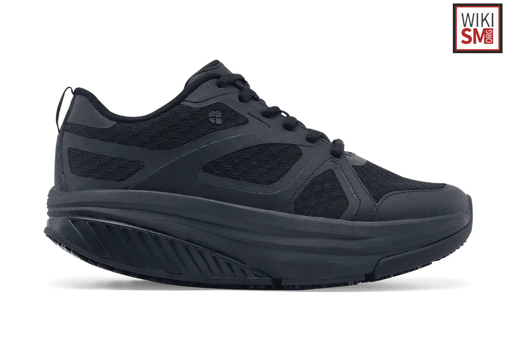
- Shoe Modification
- Optimal shoe has deep toe box (decrease contact on dorsal osteophytes), stiff sole with limited movement of 1st MTPJ
- Shoe rocker sole may decrease movement by causing a rolling transition between heel-strike and toe-off.
- Wide toe shoe
- Avoid high heels
- Foot Orthotics
- Goal: stiffen shoe, limit dorsiflexion of 1st MTPJ[12]
- Footplate made of spring-steel or carbon fibre
- Extended shank
- Morton's Extension: limits movement at the hallux, better tolerated by active patients
- Activity modification
- Avoidance of activities that cause repetitive dorsiflexion of the first MTP
- This includes running, jumping, and traveling upstairs
- Corticosteroid Injection
- Commonly used, likely beneficial with less severe arthritis
- When combined with manipulation under anesthesia, appears to help relieve symptoms and delay surgery in grade 1 and 2 disease, but not grade 3/4[13]
- Hyaluronic Acid
- RCT of 151 patients failed to show any reduction in pain at 3 months compared to placebo[14]
Surgical Management
- Indications
- When conservative management fails
- Technique
- Joint debridement (Cheilectomy)
- MTPJ Arthrodesis
- Moberg osteotomy
- Watermann osteotomy
- Youngswick osteotomy
- Keller resection arthroplasty
- MTPJ arthroplasty
- Salvage arthrodesis
Rehab and Return to Play
Rehabilitation
- Needs to be updated
Return to Play/ Work
- Needs to be updated
Complications & Prognosis
Prognosis
- Nonsurgical management
- One study of 700 patients reported a success rate of 55%[15]
Complications
- Chronic foot pain
See Also
- Internal
- External
- Sports Medicine Review Foot Pain: https://www.sportsmedreview.com/by-joint/foot/
References
- ↑ van Saase, JL, van Romunde, LK, Cats, A, Vandenbroucke, JP, Valkenburg, HA. Epidemiology of osteoarthritis: Zoetermeer survey. Comparison of radiological osteoarthritis in a Dutch population with that in 10 other populations. Ann Rheum Dis. 1989;48(4):271–280.
- ↑ Roddy, E, Thomas, MJ, Marshall, M. The population prevalence of symptomatic radiographic foot osteoarthritis in community-dwelling older adults: cross-sectional findings from the clinical assessment study of the foot. Ann Rheum Dis. 2015;74(1):156–163.
- ↑ Coughlin MJ. Conditions of the forefoot. In: DeLee J, Drez D, eds. Orthopaedic sports medicine: principles and practice. Philadelphia: WB Saunders, 1994; P221e44.
- ↑ Shurnas P, Coughlin M. Arthritic conditions of the foot. In: Surgery of the foot and ankle. Philadelphia: Elsevier, 2007; 867e909.
- ↑ McMaster MJ. The pathogenesis of hallux rigidus. J Bone Joint Surg Br 1978; 60B: 82e7.
- ↑ Coughlin, MJ, Shurnas, PS. Hallux rigidus. Grading and long-term results of operative treatment. J Bone Joint Surg Am. 2003;85-A (11):2072–2088.
- ↑ Coughlin MJ, Shurnas PS. Hallux rigidus: demographics, etiology, and radiographic assessment. Foot Ankle Int 2003; 24: 731e43.
- ↑ Case courtesy of The Radswiki, Radiopaedia.org, rID: 11470
- ↑ Kunnasegaran R, Thevendran G. Hallux rigidus: nonoperative treatment and orthotics. Foot Ankle Clin 2015; 20: 401e12.
- ↑ Yee G, Lau J. Current concepts review: hallux rigidus. Foot Ankle Int 2008; 29: 637e46.
- ↑ Hamid KS, Parekh SG. Clinical presentation and management of hallux rigidus. Foot Ankle Clin 2015; 20: 391e9.
- ↑ Sammarco VJ, Nichols R. Orthotic management for disorders of the hallux. Foot Ankle Clin 2005; 10: 191e209.
- ↑ Solan MC, Calder JD, Bendall SP. Manipulation and injection for hallux rigidus. Is it worthwhile? J Bone Joint Surg Br 2001; 83: 706e8.
- ↑ Munteanu SE, Zammit GV, Menz HB, et al. Effectiveness of intraarticular hyaluronan (Synvisc, hylan G-F 20) for the treatment of first metatarsophalangeal joint osteoarthritis: a randomised placebo-controlled trial. Ann Rheum Dis 2011; 70: 1838e41.
- ↑ Grady JF, Axe TM, Zager EJ, Sheldon LA. A retrospective analysis of 772 patients with hallux limitus. J Am Podiatr Med Assoc 2002; 92: 102e8.
Created by:
John Kiel on 20 June 2019 21:15:13
Authors:
Last edited:
26 November 2025 16:02:08
Categories: