Foot Drop
(Redirected from Peroneal Nerve Injury)
Other Names
- Deep Peroneal Nerve Injury
- Superficial Peroneal Nerve Injury
- Common Peroneal Nerve Injury
- Peroneal Neuropathy
- Peroneal nerve compromise
- Peroneal Neuropraxia
- Foot drop
- Peroneal Nerve Palsy
Background
- This page refers to peripheral neuropathies of, referred to as peroneal nerve injury (PNI), or more commonly 'Foot Drop'
- Common Peroneal Nerve (CPN)
- Deep Peroneal Nerve (DPN)
- Superficial Peroneal Nerve (SPN)
History
- First described in 1838 (need citation)
- PNI following ankle sprain first described by Hyslop in 1941[1]
Epidemiology
- One study found Peroneal Nerve Injuries were the most common lower extremity neuropathy in athletes[2]
- Of 303 patients preventing with foot drop, 31% were found to be due to peroneal neuropathy[3]
- Nerve involvement
- Krivickas et al found the majority of cases were CPN or proximal DPN[2]
Introduction


General
- Defined as weakness or inability to dorsiflex the ankle, most commonly due to dysfunction of the neural pathway to the dorsiflexor muscles, such as L5 radiculopathy or peroneal nerve injury
- Present with a high-stepping gait and may compensate with abnormal walking patterns to avoid tripping, often accompanied by muscle weakness and sometimes sensory deficits
- Diagnosis relies on a thorough history, physical examination, and localization of the lesion using electrodiagnostic studies and imaging modalities
- Management includes addressing the underlying etiology, physical therapy, use of ankle-foot orthoses
- See also: Neuropathies Main
Etiology: Traumatic
- Knee Dislocation
- CPN Palsy is seen in 5-40% of knee dislocations[6]
- Proximal Fibula Fracture
- Proximal Tibia Fracture
- Tibial Plateau Fracture
- Estimated 1% incidence (need citation)
- ACL Injury, less commonly other ligamentous injuries
- Ankle Fracture
- Ankle Sprain
- Direct trauma
- Examples include direct impact, penetrating trauma, laceration
- Most commonly this is around the fibular head where the nerves are most superficial
Etiology: Iatrogenic
- Arthroscopic meniscal repair
- Realignment of the knee extensor mechanism
- Knee Arthrodesis
- Total Knee Replacement
- Treatment of knee flexion contracture
- Posterior Short Leg Splint or Short Leg Cast
- Can damage the peroneal nerve by pressure over the fibular head
- Mitigated by padding the cast or splint in the area of the fibular head
- Tight compression or bandage
- Position during anesthesia or surgery
Etiology: Other
- Mass effect
- Ganglion Cyst or Bakers Cyst is a common mass lesion associated with PNI, specifically the CPN[9]
- Bakers Cyst was the most common cause of PNI by mass effect in a study by Kim et al[10]
- Tumors in order of decreasing frequency, included schwannoma, neurofibroma, osteochondroma, neurogenic sarcoma, focal hypertrophic neuropathy, desmoid tumor, and glomus tumor
- Knee Osteoarthritis
- Several case reports, including one with varus laxity[11]
- Habitual leg crossing
- Prolonged bed rest
Anatomy of the Common Peroneal Nerve and branches
- Common Peroneal Nerve (CPN)
- One of two branches off the Sciatic Nerve (the other is the Tibial Nerve)
- Nerve Roots: L4 through S1
- Bifurcation occurs above popliteal fossa, CPN then travels across lateral gastroc
- Innervation: cutaneous aspect of lateral leg below knee
- Dives deep to fibular head, bifurcates into DPN and SPN
- Deep Peroneal Nerve (DPN)[12]
- Motor innervation: extensor muscles of ankle, foot
- Sensory innervation: web space between the first and second toes
- Superficial Peroneal Nerve (SPN)
- Motor innervation: ankle eversion, plantarflexion
- Sensory innervation: skin of the lateral leg, dorsum of the foot and toes, sparing the small area between the first two toes
- 15-28% percent of patients have an accessory peroneal nerve that branches off the superficial peroneal to supply the extensor digitorum brevis
Risk Factors
- Sports
- Biomechanical
- Prolonged squatting[13]
- Other
- Systemic Illness
- Diabetes Mellitus
- Motor Neuron Disease
- Anorexia Nervsosa (due to loss of subcutaneous fat leading to nerve compression)
Differential Diagnosis
Differential Diagnosis Leg Pain
- Fractures & Dislocations
- Muscle and Tendon Injuries
- Neurological
- Vascular
- Other
- Pediatric Considerations
- Tibial Tubercle Avulsion Fracture
- Tibial Tuberosity Apophysitis
- Toddlers Fracture (Tibial Shaft Fracture)
Differential Diagnosis Neuropathy
- L5 Radiculopathy
- Lumbosacral Plexopathy
- Sciatic Nerve Injury
- Peroneal Nerve Injury
Clinical Features


History
- Need to characterize nature of the presentation including acuity (chronic vs acute), any history of trauma, etc
- Inquire about prolonged leg crossing, compressive positions, history of back pain, recent trauma
- Typically complain of lateral lower limb and dorsal foot pain
- Concurrent low back pain or posterolateral thigh pain suggests L5 radiculopathy
- Pain usually precedes sensory changes in a similar distribution
- Patient may complain of foot drop as the first manifestation of this disorder
- Can describe difficulty walking, tripping, slapping sound of foot
- Much less commonly central causes such as brain or spinal cord lesions may have additional features
Physical Exam: Physical Exam Leg
- Hallmark finding is weakness in ankle dorsiflexion
- Weakness of tibialis anterior, extensor hallucis longus, extensor digitorum longus
- Weakness in toe extension, eversion of foot may be present
- Careful sensory examination can localize the lesion
- Sensory deficity is typically present over lateral leg, dorsal foot
- PNI should have sensory and motor deficits limited to the calf, ankle and foot
- Involvement of the knee or anything proximal suggests sciatic nerve, lumbosacral radiculopathy
- Gait analysis usually reveals a high stepping gait if the patient is compensating
- Upper motor neuron signs are typically absent
Common Peroneal Nerve
- Sensory: Lateral calf and dorsum of foot (sparing lateral and plantar foot)
- Weakness: Ankle dorsiflexion and eversion, toe extension
Deep Peroneal Nerve
- Sensory: Area between great and second toes
- Weakness: Ankle dorsiflexion, partial eversion > inversion, toe extension
Superficial Peroneal Nerve
- Sensory: Lateral calf and dorsum of foot (sparing lateral foot)
- Weakness: Ankle eversion
Special Tests
- Tinels Test: tapping around the fibular head may reproduce symptoms
- Babinski Reflex: should be normal in patients with peripheral foot drop
Evaluation
General Approach
- Guided by clinical localization and is typically used to identify the underlying etiology
- Especially when the diagnosis is unclear or when a structural lesion is suspected
Radiographs
- Standard Radiographs Knee, Standard Radiographs Ankle
- Typically normal in the absence of trauma
- Evaluate for fracture, mass lesion, arthritis
MRI
- Need minimum of 3T MRI to view peroneal nerve
- Knee/ Ankle[21]
- Consider MRI knee in all cases since intraneural ganglia may be the most common cause[22]
- Look for bony lesion, neural ganglia
- Lumbar[23]
- Should be obtained if a lumbar or central etiology is suspected
- Look for evidence of L5 radiculopathy
Ultrasound
- Assess fibular head[24]
- Can detect nerve enlargment, masses or focal entrapment
- Dynamic evaluation can also help localize lesions in uncertain cases
EMG/NCS
- Helpful with
- confirm diagnosis of peroneal neuropathy
- Exclude alternative diagnoses
- Prognosticate
- Evaluation should include
- Motor nerve conduction studies of the peroneal nerve, tibial nerve
- Sensory nerve conduction studies of the sural, superficial peroneal nerves
- Findings
- If demyelination, focal slowing or conduction block
- If axon loss, compound muscle action potential amplitudes will be decreased
- If abnormal, must examine nerves supplied by L5 nerve root but not by peroneal nerve
- Includes Tibialis Posterior, medial gastrocnemius,
- Expand differential of radiculopathy, lumbosacral plexopathy, sciatic neuropathy
Classification
The Sunderland Classification of Peripheral Nerve Injuries
- Grade 1: Conduction block +/- segmental demyelination
- Grade 2: Axon discontinuity with intact endoneurium
- Grade 3: Axon and endoneurium discontinuity with intact perineurium
- Grade 4: Axon, endoneurium and perineurium discontinuity with intact epineurium
- Grade 5: Nerve discontinuity
- Grade 6: Mixed nerve injury
Management

Nonoperative
- Indications
- Vast majority of cases
- No clearly identified mass or lesion which could be surgically excised
Analgesia
- General
- Neuropathic pain is challenging to treat
- Consider the following agents which should be individualized to the patient
- Topical lidocaine
- Capsaicin
- Selective serotonin reuptake inhibitors
- Antiepileptics
- Opioids
- μ-receptor agonists
- Heat Therapy
- If any sensory loss, be careful to avoid thermal damage
- Ice Therapy
- If any sensory loss, be careful to avoid thermal damage
- Case report suggested ice applied to the fibular head lead to a peroneal nerve palsy[26]
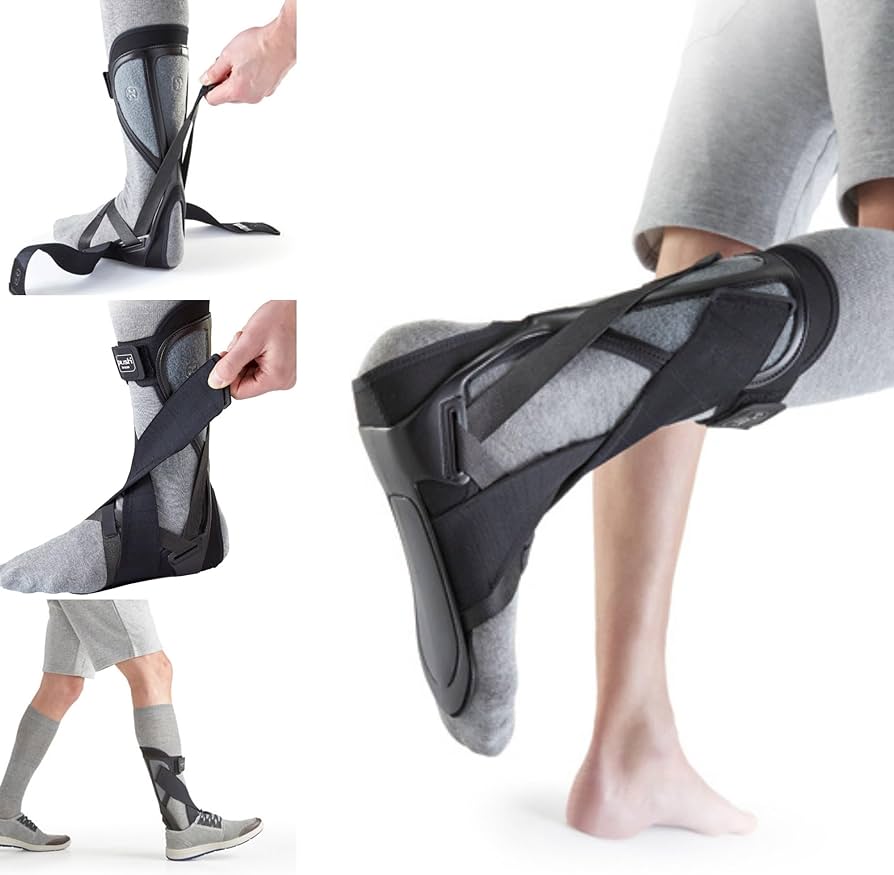
Orthotics
- Ankle Foot Orthosis
- Necessary if unable to dorsiflex ankle
- Seen with proximal deep peroneal neuropathy, common peroneal nerve
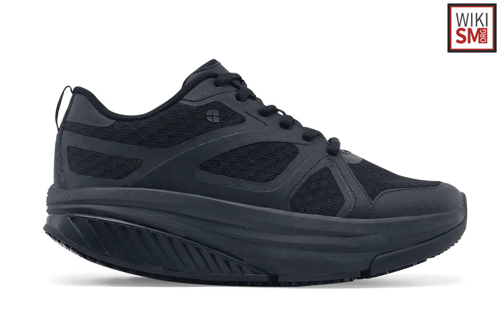
- Rocker Bottom Shoe
- Consider if weakness of the toe extensors only (distal deep peroneal neuropathy)
- Can be used to optimize gait, reduce energy for ambulation
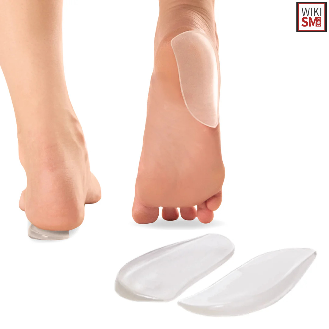
- Lateral wedge
- Consider if isolated superficial peroneal nerve palsy
- Prevent supination from weak eversion
Other Modalities
- Iontophoresis
- Physical Therapy
- Useful in subtle peroneal nerve injuries to help with recovery
- If complete nerve transection or axonal loss, strengthening is not helpful, PROM may
- Maintain ROM important to avoid heel cord contracture
- Peroneal Nerve Stimulator
- Require an intact functioning peroneal nerve
- Not useful in patients with peripheral nerve injury.
- Wound checks
- Patients with sensory loss should check their feet daily to prevent progression of ulcers
Operative
- Indication
- Mass or lesion causing neuropathy
- Refractory pain
- Inability to ambulate with AFO due to contracture
- Technique
- Surgical repair or graft
- Tibialis posterior tendon transfer (to restore active dorsiflexion)
- Neurolysis (for chronic pain)
Rehab and Return to Play
Rehabilitation
- Strength training shows variable efficacy depending on the underlying condition
- Progressive resistance exercises for ankle dorsiflexors, gait training, and endurance activities.
- Stretching/ ROM
- Important to stretch daily to prevent contracture, maintain ambulatory status
- Patient often cant tolerate bracing if contracture develops
Return to Play/ Work
- Initial phase (0-6 weeks)[27]
- Device fitting (AFO or FES) with 8 dose-matched physical therapy sessions focusing on gait mechanics, strength, and endurance
- Progressive rehabilitation (6-12 weeks)[28]
- Advancement of strengthening exercises, treadmill training with FES if available
- Functional activities with objective monitoring of gait parameters
- Advanced functional training (12-24 weeks)
- Sport-specific movements, agility drills, and endurance activities
- Monitoring for fatigue-related gait deterioration
- Return to sport criteria[29]
- Ankle dorsiflexion to ≥15 degrees at terminal swing
- Normalized step length asymmetry
- Maintenance of gait speed during endurance activities
- Adequate balance on stabilometric testing
- Patient-reported functional satisfaction
Prognosis and Complications
Prognosis
- Prognosis varies widely and is dependent on etiology
- Following Knee Dislocation
- Complete palsy holds a worse prognosis than incomplete palsy[30]
- Following Total Knee Arthroplasty
- Park et al found 62% of patients had maximal recovery, 38% had complete recovery at 12 months[31]
- Those requiring surgical management
- Predictors of good outcome
- Patients with muscle strength grade 2/5 or above are significantly more likely to achieve greater recovery[34]
- Patients with intervention within first 6 weeks are 6 times more likely to demonstrate greater recovery[35]
- Younger patients, radicular pain and early postoperative recovery are also good predictors of better outcomes
Complications
- Physical dysfunction
- Mobility impariment
- Fall risk, injury risk
- Increased due to impaired gait mechanics[36]
- Ankle contracture
- Equinovarus foot deformity
- Need for surgery
- Chronic disability
- Approximately 20% of patients require long term use of ankle foot orthosis or other assist devices
- Gait abnormalities
- Patient dissatisfaction
- This is especially true when foot drop is a complication of spinal surgery[37]
See Also
References
- ↑ Hyslop G. Injuries to the deep and superficial peroneal nerves complicating ankle sprain. Am J Surg. 1941;11(2):436–8.
- ↑ 2.0 2.1 2.2 Krivickas, Lisa S., and Asa J. Wilbourn. "Peripheral nerve injuries in athletes: a case series of over 200 injuries." Seminars in neurology. Vol. 20. No. 02. Copyright© 2000 by Thieme Medical Publishers, Inc., 333 Seventh Avenue, New York, NY 10001, USA. Tel.:+ 1 (212) 584-4662, 2000.
- ↑ Langenhove M, Pollefliet A, Vanderstraeten G. A retrospective electrodiagnostic evaluation of footdrop in 303 patients. Electromyogr Clin Neurophysiol. 1989;29:145–52.
- ↑ Harris, Connie, et al. "Refractory venous leg ulcers: observational evaluation of innovative new technology." International Wound Journal 14.6 (2017): 1100-1107.
- ↑ Image courtesy of teachmeanatomy.info
- ↑ Robertson A, Nutton RW, Keating JF (2006) Dislocation of the knee. J Bone Joint Surg Br 88:706–711
- ↑ McCrory P, Bell S, Bradshaw C. Nerve entrapments of the lower leg. Sports Med. 2002;32(6):371–91.
- ↑ Nitz AJ, Dobner JJ, Kersey D. Nerve injury and Grades II and II ankle sprains. Am J Sports Med. 1985;13(3):177–82.
- ↑ Spinner RJ, Atkinson JL, Tiel RL. Peroneal intraneural ganglia: the importance of the articular branch. A unifying theory. J Neurosurg. 2003;99:330–43.
- ↑ Kim DH, Murovic JA, Teil RL, Kline DG. Management and outcomes in 318 operative common peroneal nerve lesions at the LSU Health Sciences Center. Neurosurgery. 2004;54:1421–9.
- ↑ Fetzer GB, Prather H, Gelberman RH, Clohisy JC. Progressive peroneal nerve palsy in a varus athritic knee. JBJS. 2004;86-A(7):1538–40.
- ↑ Jenkins DB. The Leg. In: Hollinshead’s functional anatomy of the limbs and back. 8th ed. Philadelphia: WB Saunders; 2002. pp. 327–50.
- ↑ Togrol E. Bilateral peroneal nerve palsy induced by prolonged squatting. Mil Med. 2000;165(3):240–2.
- ↑ Katirji MB, Wilbourn AJ. Common peroneal mononeuropathy: a clinical and electrophysiologic study of 116 lesions. Neurology. 1988;38:1723–8
- ↑ Fukuda H. Bilateral peroneal nerve palsy caused by intermittent pneumatic compression. Intern Med. 2006;45(2):93–4.
- ↑ Lu, Hui, et al. "A rapidly progressive foot drop caused by the posttraumatic Intraneural ganglion cyst of the deep peroneal nerve." BMC musculoskeletal disorders 19.1 (2018): 298.
- ↑ Ricarte, Irapuá Ferreira, et al. "Acute foot drop syndrome mimicking peroneal nerve injury: an atypical presentation of ischemic stroke." Journal of Stroke and Cerebrovascular Diseases 23.5 (2014): 1229-1231.
- ↑ Bueno-Gracia, Elena, et al. "Neurodynamic test of the peroneal nerve: Study of the normal response in asymptomatic subjects." Musculoskeletal Science and Practice 43 (2019): 117-121.
- ↑ Swiatek, Peter, et al. "Examining the anatomy, pathophysiology, and clinical presentation of lower extremity neurologic deficits: a spine surgeon's guide to foot drop." JAAOS-Journal of the American Academy of Orthopaedic Surgeons 33.18 (2025): e1060-e1071.
- ↑ Işık, Semra, et al. "Bilateral central foot drop in a pediatric patient." Pediatric Neurosurgery 52.1 (2016): 62-66.
- ↑ Park, Se-Heum, Hwan-Kwon Do, and Geun-Yeol Jo. "Compressive peroneal neuropathy by an intraneural ganglion cyst combined with L5 radiculopathy: A case report." Medicine 98.44 (2019): e17865.
- ↑ Kim JY, Ihn YK, Kim JS, Chun KA, Sung MS, Cho KH. Non-traumatic peroneal nerve palsy: MRI findings. Clin Radiol. 2007;62:58–64.
- ↑ Macki, Mohamed, et al. "Clinching the cause: a review of foot drop secondary to lumbar degenerative diseases." Journal of the neurological sciences 395 (2018): 126-130.
- ↑ Grant, Thomas H., et al. "Sonographic evaluation of common peroneal neuropathy in patients with foot drop." Journal of Ultrasound in Medicine 34.4 (2015): 705-711.
- ↑ Felici, Nicola, et al. "Common peroneal nerve injuries at the knee: outcomes of nerve repair." PRRS 1.1 (2022): 6-13.
- ↑ Moeller JL, Munroe J, McKeag DB. Cryotherapy induced common peroneal nerve palsy. Clin J Sports Med. 1997;7:212–6.
- ↑ Kluding, Patricia M., et al. "Foot drop stimulation versus ankle foot orthosis after stroke: 30-week outcomes." Stroke 44.6 (2013): 1660-1669.
- ↑ David, Romain, et al. "A 6-month home-based functional electrical stimulation program for foot drop in a post-stroke patient: Considerations on a time course analysis of walking performance." International Journal of Environmental Research and Public Health 19.15 (2022): 9204.
- ↑ Orunoğlu, Merdan, et al. "Outcomes of Posterior Tibial Tendon Transfer in Patients With Foot Drop: A Case Series of 11 Patients." World Neurosurgery (2025): 124415.
- ↑ O'Malley MP, Pareek A, Reardon P, Krych A, Stuart MJ, Levy BA. Treatment of Peroneal Nerve Injuries in the Multiligament Injured/Dislocated Knee. J Knee Surg. 2016 May;29(4):287-92.
- ↑ Park, Jai Hyung, et al. "Common peroneal nerve palsy following total knee arthroplasty: prognostic factors and course of recovery." The Journal of arthroplasty 28.9 (2013): 1538-1542.
- ↑ Poage, Chad, Charles Roth, and Brandon Scott. "Peroneal nerve palsy: evaluation and management." JAAOS-Journal of the American Academy of Orthopaedic Surgeons 24.1 (2016): 1-10.
- ↑ Liu, Kun, et al. "Foot drop caused by lumbar degenerative disease: clinical features, prognostic factors of surgical outcome and clinical stage." PLoS One 8.11 (2013): e80375.
- ↑ Baig Mirza, Asfand, et al. "Prognostic factors and surgical outcomes of foot drop secondary to lumbar degenerative disease: A systematic review and Individual patient data meta-analysis." European Spine Journal (2025): 1-12.
- ↑ Tanaka, Jun, et al. "Drop foot due to lumbar degenerative disease: painless drop foot is difficult to recover." Clinical Neurology and Neurosurgery 206 (2021): 106696.
- ↑ Dwivedi, Nishant, et al. "Surgical treatment of foot drop: patient evaluation and peripheral nerve treatment options." Orthopedic Clinics 53.2 (2022): 223-234.
- ↑ Swiatek, Peter, et al. "Examining the anatomy, pathophysiology, and clinical presentation of lower extremity neurologic deficits: a spine surgeon's guide to foot drop." JAAOS-Journal of the American Academy of Orthopaedic Surgeons 33.18 (2025): e1060-e1071.
Created by:
John Kiel on 7 July 2019 07:26:38
Authors:
Last edited:
4 December 2025 18:37:28
Categories: