Meniscus
(Redirected from Lateral meniscus)
Description
-
![(A) Basic anatomy of the knee. (B) Cross-sectional diagram of the meniscus.[1]](/w/images/thumb/f/fc/Meniscus_anatomy_cross_section.jpeg/120px-Meniscus_anatomy_cross_section.jpeg) (A) Basic anatomy of the knee. (B) Cross-sectional diagram of the meniscus.[1]
(A) Basic anatomy of the knee. (B) Cross-sectional diagram of the meniscus.[1] -
![(A) Anatomy of the meniscus viewed from above (B) Axial view of a right tibial plateau showing sections of the meniscus and their relationship to the cruciate ligaments. AL, anterior horn lateral meniscus; AM, anterior horn medial meniscus; PCL, posterior cruciate ligament; PL, posterior horn lateral meniscus; PM, posterior horn medial meniscus.[2]](/w/images/thumb/2/23/Meniscus_anatomy.jpeg/84px-Meniscus_anatomy.jpeg) (A) Anatomy of the meniscus viewed from above (B) Axial view of a right tibial plateau showing sections of the meniscus and their relationship to the cruciate ligaments. AL, anterior horn lateral meniscus; AM, anterior horn medial meniscus; PCL, posterior cruciate ligament; PL, posterior horn lateral meniscus; PM, posterior horn medial meniscus.[2]
(A) Anatomy of the meniscus viewed from above (B) Axial view of a right tibial plateau showing sections of the meniscus and their relationship to the cruciate ligaments. AL, anterior horn lateral meniscus; AM, anterior horn medial meniscus; PCL, posterior cruciate ligament; PL, posterior horn lateral meniscus; PM, posterior horn medial meniscus.[2] -
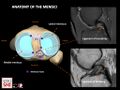
![Axial image (C) shows the normal appearance of the menisci, location of sagittal slices (lines 1. and 2.) in B, C and D. Sagittal images show the typical bow-tie configuration of the meniscal body corresponding to line 1 (B) and the opposing triangles of the horns corresponding to line 2 (C and D). Moreover, the horns of the LM are similar in size and shape (C), whereas the posterior horn of the MM is larger than the anterior horn (D)[3]](/w/images/thumb/9/93/MRI_meniscus_anatomy.jpeg/114px-MRI_meniscus_anatomy.jpeg) Axial image (C) shows the normal appearance of the menisci, location of sagittal slices (lines 1. and 2.) in B, C and D. Sagittal images show the typical bow-tie configuration of the meniscal body corresponding to line 1 (B) and the opposing triangles of the horns corresponding to line 2 (C and D). Moreover, the horns of the LM are similar in size and shape (C), whereas the posterior horn of the MM is larger than the anterior horn (D)[3]
Axial image (C) shows the normal appearance of the menisci, location of sagittal slices (lines 1. and 2.) in B, C and D. Sagittal images show the typical bow-tie configuration of the meniscal body corresponding to line 1 (B) and the opposing triangles of the horns corresponding to line 2 (C and D). Moreover, the horns of the LM are similar in size and shape (C), whereas the posterior horn of the MM is larger than the anterior horn (D)[3] -
 Anatomy of the menisci (right knee). Anatomy of the meniscofemoral ligaments. ACL: anterior cruziate ligament. PCL: posterior cruziate ligament.
 -
![A variety of meniscal tears are illustrated.[4]]]](/w/images/thumb/c/c7/Meniscus_tear_patterns.jpeg/120px-Meniscus_tear_patterns.jpeg) A variety of meniscal tears are illustrated.[4]]]
A variety of meniscal tears are illustrated.[4]]]
![(A) Basic anatomy of the knee. (B) Cross-sectional diagram of the meniscus.[1]](/File:Meniscus_anatomy_cross_section.jpeg)
![(A) Anatomy of the meniscus viewed from above (B) Axial view of a right tibial plateau showing sections of the meniscus and their relationship to the cruciate ligaments. AL, anterior horn lateral meniscus; AM, anterior horn medial meniscus; PCL, posterior cruciate ligament; PL, posterior horn lateral meniscus; PM, posterior horn medial meniscus.[2]](/File:Meniscus_anatomy.jpeg)
![Axial image (C) shows the normal appearance of the menisci, location of sagittal slices (lines 1. and 2.) in B, C and D. Sagittal images show the typical bow-tie configuration of the meniscal body corresponding to line 1 (B) and the opposing triangles of the horns corresponding to line 2 (C and D). Moreover, the horns of the LM are similar in size and shape (C), whereas the posterior horn of the MM is larger than the anterior horn (D)[3]](/File:MRI_meniscus_anatomy.jpeg)

![A variety of meniscal tears are illustrated.[4]]]](/File:Meniscus_tear_patterns.jpeg)


Other Names
- Medial Meniscus
- Lateral Meniscus
- Menisci
- Knee meniscus
- Meniscal cartilage
- Semilunar cartilage
- Fibrocartilage of the knee
- Tibiofemoral meniscus
General
- Crescent-shaped wedges of fibrocartilage oriented circumferentially
- Positioned between the tibial plateaus and the femoral condyles in the medial and lateral compartments
- Menisci possess collagen fibers oriented circumferentially
- These circumferential fibers are bound by radially oriented fibers
Structure
- Made primarily of type I collagen bundles
- Circumferential and radially oriented to help prevent tearing[5]
- Contain types I and II neuroreceptors
- Possible proprioceptive and mechanoreceptive capacities
Medial Meniscus
- Larger of the two menisci
- C-shaped and covers about 50% of the medial tibial plateau
- Posterior horn is larger than anterior horn
- Bony attachments to the anterior and posterior horns at the meniscal root
- Peripherally, attaches to the joint capsule, deep fibers of the MCL
- Providers anteroposterior stability to the knee
- Stronger attachment to joint capsule, less mobile, more susceptible to separation and tears
Lateral Meniscus
- More circular than the medial meniscus
- Covers about 70% of the lateral tibial plateau
- Posterior and anterior horns are similar in size
- Anterior attachment next to the ACL
- Posterior attachment behind intercondylar eminence, anterior to the attachment of the medial meniscus
- It also has attachment to the PCL via meniscofemoral ligaments, popliteomeniscal fasculi and the ligament of Wrisberg


Discoid Meniscus
- Congenital variant with abnormal morphology, can create innate instability of the lateral meniscus
- Thicker, poor tissue quality, less vascularity than a normal meniscus
- Prone to tears which can be symptomatic or asymptomatic
Watanabe Classification for Discoid Meniscus[10]
- Type I/ Wrisberg
- Least common, meniscotibial attachment of lateral meniscus is absent
- Most unstable variation of discoid meniscus[8]
- Type II/ complete
- Most common type, meniscus covers the entire tibial plateau
- Typically thickened and hypertrophic
- Type III/ incomplete
- More common than Type I, less common than Type II
- partially covers tibial plateau, also thickened
- Normal tibial attachment
Primary function[11]
- Menisci transmits 50% of joint compressive forces in full extension
- Approximately 85% of the load in 90° of flexion
Contributes To
- Protect articular cartilage
- Shock absorption
- Augment lubrication
- Rotation of the opposing articular surfaces
- Joint nutrition
- Tibiofemoral joint stability
- Joint congruency
- Proprioception[12]
Vascular Supply


- Distribution
- The blood supply to the meniscus begins in the periphery which allows these regions to have the best healing
- The outer third is considered red due to the good blood supply
- The inner two thirds is considered white and is avascular, relies on synovial fluid for nutrition
- Vascular classification
- Based on the side of the tear they can be classified as white-white, red-red, or white-red.
- Those tears in the white-white are avascular and typically do not heal.
- Originates from geniculate arteries: superior, inferior, medial and lateral
- Perimeniscal capillary plexus originating in the knee’s capsular and synovial tissues
Innervation
- Medial Meniscus innervated by articular branches of:
- Saphenous Nerve
- Nerve to Vastus Medialis
- Obturator Nerve (posterior articular branch)
- Tibial Nerve
- Lateral Meniscus innervated by articular branches of:
- Common Fibular Nerve
- Tibial Nerve (posterior articular branches)
- Nerve to Vastus Lateralis
Clinical Significance
See Also
References
- ↑ Li, Hao, et al. "Meniscal regenerative scaffolds based on biopolymers and polymers: recent status and applications." Frontiers in Cell and Developmental Biology 9 (2021): 661802.
- ↑ Fox, Alice JS, Asheesh Bedi, and Scott A. Rodeo. "The basic science of human knee menisci: structure, composition, and function." Sports health 4.4 (2012): 340-351.
- ↑ Anghel, A-I., G. S. Toma, and D. Cuzino. "A pictorial review of knee meniscus-from anatomy to complex tears." European Congress of Radiology-ECR 2024, 2024.
- ↑ Case courtesy of Matt Skalski, Radiopaedia.org, rID: 55569
- ↑ 5.0 5.1 Torres, Stephen J., Jason E. Hsu, and Robert L. Mauck. "Meniscal anatomy." Meniscal Injuries: Management and Surgical Techniques (2014): 1-7.
- ↑ Vadodaria, Ketankumar, et al. "Materials and structures used in meniscus repair and regeneration: a review." Biomedicine 9.1 (2019): 2.
- ↑ Fox, Alice JS, et al. "The human meniscus: a review of anatomy, function, injury, and advances in treatment." Clinical anatomy 28.2 (2015): 269-287.
- ↑ 8.0 8.1 Hirschmann, M. T., and N. F. Friederich. "Classification: discoid meniscus, traumatic lesions." The Meniscus (2010): 241-246.
- ↑ Prescott, Jeffrey William. Computer-assisted discovery and characterization of imaging biomarkers for disease diagnosis and treatment planning. Diss. The Ohio State University, 2010.
- ↑ Chambers, Henry G., and Reid C. Chambers. "The natural history of meniscus tears." Journal of Pediatric Orthopaedics 39 (2019): S53-S55.
- ↑ Lee SJ, Aadalen KJ, Malaviya P, et al. Tibiofemoral contact mechanics after serial medial meniscectomies in the human cadavcadaveric knee. Am J Sports Med 2006;34(8):1334-1344.
- ↑ Zimny ML, Albright DJ, Dabezies E. Mechanoreceptors in the human medial meniscus. Acta Anat Basel 1988;133(1):35-40.
- ↑ van Schie, Peter, et al. "Intra-operative assessment of the vascularisation of a cross section of the meniscus using near-infrared fluorescence imaging." Knee Surgery, Sports Traumatology, Arthroscopy (2021): 1-10.
- ↑ Mameri, Enzo S., et al. "Review of meniscus anatomy and biomechanics." Current reviews in musculoskeletal medicine 15.5 (2022): 323-335.