Ligament of Wrisberg
Description
-
![The ligament of Wrisberg on sagittal proton density-weighted imaging[1]](/w/images/thumb/3/31/Ligament_of_Wrisberg_MRI.jpeg/120px-Ligament_of_Wrisberg_MRI.jpeg) The ligament of Wrisberg on sagittal proton density-weighted imaging[1]
The ligament of Wrisberg on sagittal proton density-weighted imaging[1] -
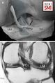 Computer drawing (posterior view of the right knee) demonstrating the meniscofemoral ligaments (MFLs) of Humphry and Wrisberg, and the pseudotear zone (P). b Corresponding coronal T1-weighted spin-echo MR image (600/14). PCL posterior cruciate ligament, MM medial meniscus, LM lateral meniscus
 -
![MRI of wrisberg annotated[2]](/w/images/thumb/0/0f/MRI_ligament_of_wrisberg.jpeg/120px-MRI_ligament_of_wrisberg.jpeg) MRI of wrisberg annotated[2]
MRI of wrisberg annotated[2] -
![Anatomy of the knee including Ligament of Wrisberg[3]](/w/images/thumb/d/df/Anatomy_knee_ligament_of_wrisberg.jpeg/120px-Anatomy_knee_ligament_of_wrisberg.jpeg) Anatomy of the knee including Ligament of Wrisberg[3]
Anatomy of the knee including Ligament of Wrisberg[3]
![The ligament of Wrisberg on sagittal proton density-weighted imaging[1]](/File:Ligament_of_Wrisberg_MRI.jpeg)

![MRI of wrisberg annotated[2]](/File:MRI_ligament_of_wrisberg.jpeg)
![Anatomy of the knee including Ligament of Wrisberg[3]](/File:Anatomy_knee_ligament_of_wrisberg.jpeg)


Name
- Posterior meniscofemoral ligament (PMFL)
- Wrisberg ligament
- Meniscofemoral ligament of Wrisberg
- Posterior meniscal ligament
- Meniscofemoral ligament
General
- Small ligament in the knee joint, part of the meniscofemoral ligaments
- Arises from posterior horn of the lateral meniscus
- Passes superomedial and behind the posterior cruciate ligament
- Inserts on the lateral surface of the medial femoral condyle
Function
- Contributes to stabilization of the posterolateral part of the lateral meniscus
- Helps pull lateral meniscus anteromedially during flexion
- Improves congruence of lateral compartment, decreasing contact pressure
- Protective role of the PCL
Anatomic Variance
- Reportedly present in approximately 65% of patients[6]
MRI Features
- Best visualized in coronal and sagittal planes
Clinical Significance
- Meniscofemoral ligament tear
- May be mistaken for meniscus tear, loose body
See Also
References
- ↑ 유혜진, et al. "수술 전 반월연골: 피해야 할 함정들." Journal of the Korean Society of Radiology 83.3 (2022): 582-596.
- ↑ Case courtesy of Ayush Goel, Radiopaedia.org, rID: 28390
- ↑ Image courtesy of musculoskeletalkey.com
- ↑ Image courtesy of https://www.kneeguru.co.uk/
- ↑ Deckey, David G., et al. "Prevalence, biomechanics, and pathologies of the meniscofemoral ligaments: a systematic review." Arthroscopy, Sports Medicine, and Rehabilitation 3.6 (2021): e2093-e2101.
- ↑ Mohankumar, Rakesh, Lawrence M. White, and Ali Naraghi. "Pitfalls and pearls in MRI of the knee." American Journal of Roentgenology 203.3 (2014): 516-530.