Acromioclavicular Joint Separation
(Redirected from Acromioclavicular Joint Dislocation)
Other Names
- Shoulder Separation
- Shoulder Sprain
- AC Sprain
- AC Joint Sprain
- AC Dislocation
- Acromioclavicular Joint Dislocation
Background
- This page refers to all traumatic injuries to the Acromioclavicular Joint including
- Sprains
- Dislocations
- Separations
History
- Needs to be updated
Epidemiology
- Thought to be under-estimated due to many mild injuries not seeking medical attention
- Represents 9% of all shoulder injuries (need citation)
- 50% of all AC dislocations occur to individuals in their 20s (need citation)
- Types 1 and 2 injuries account for most AC separations
Introduction


General
- Characterized by anterior/lateral shoulder pain with or without deformity
- Mechanism of injury is primarily by direct impact over the AC joint or lateral shoulder
- Diagnosis is clinical and confirmed with radiographs or ultrasound
- Management is generally considered nonsurgical
Etiology
- Simple falls
- High energy trauma
- Sports
- From fall or contact sport
- Arm is adducted
Anatomy of the Acromioclavicular joint
- General
- Diarthrodial joint
- Motion: primarily gliding, only 8° rotation through AC joint
- fibrocartilaginous intraarticular disc (similar to meniscus of knee)
- Stabilizers
- Joint Capsule
- Acromioclavicular Ligament (anterior-posterior stability)
- Coracoclavicular Ligaments (superior-inferior stability)
- Muscles: Deltoid, Trapezius
Associated Injuries
- Intra-articular shoulder injury (18.2%)[2]
- SLAP Lesion
- Rotator Cuff Injury
Risk Factors
- Male > Female
- Sports
- Contact and collision sports
- Skiing
- Cycling
Differential Diagnosis
- Fractures
- Proximal Humerus Fracture
- Humeral Shaft Fracture
- Clavicle Fracture
- Scapula Fracture
- First Rib Fracture (traumatic or atraumatic)
- Floating Shoulder
- Dislocations & Separations
- Arthropathies
- Muscle & Tendon Injuries
- Rotator Cuff
- Bursopathies
- Ligament Injuries
- Neuropathies
- Other
- Pediatrics
- Coracoid Avulsion Fracture
- Humeral Head Epiphysiolysis (Little League Shoulder)
Clinical Features

History
- Patient should describe some trauma
- Shoulder pain at or close to AC joint
- Deformity may or may not be present
Physical: Physical Exam Shoulder
- Most commonly, tenderness over AC joint
- Inspection may show asymmetry of shoulders or swelling at the AC joint
- Range of Motion is usually reduced due to pain acutely
- Pain with adduction of the shoulder and possibly with shoulder abduction and flexion
- Deformity (step-off) of AC joint with type III injuries and higher
- Type III and V may show instability of the lateral clavicle when depressed manually ("piano key" phenomenon)
Special Tests
- Crossover Test: shoulder flexed to 90°, passively adducted across chest, reproduces pain over the AC joint
- Resisted AC Joint Extension Test: abduction against resistance with shoulder flexed to 90°
- OBriens Test: Arm flexed to 90° and then supinated and pronated against resistance
Evaluation
-
 Grade 3 AC Joint Separation
 -
 Zanca view showing normal AC joint (right) and grade 3 separation (left)


Radiographs
- Standard Radiographs Shoulder
- May be normal in grade I/II
- More obvious in severe grade II or grade III
- Zanca View
- Sometimes referred to as the AC joint view[6]
- Beam is tilted cephalic 10 - 15°
- Provides superior AC joint evaluation
- Recommend bilateral evaluation for comparison
- Other Views
- Basmania View: scapular Y performed with cross-body adduction stress
- Stryker notch views can assess for coracoid fracture
- Weighted stress view: no longer used
- Coracoclavicular Distance
- Measures the distance between superior cortex of coracoid process and undersurface of clavicle
- Used to assess the integrity of the Coracoclavicular Ligament radiographically
- The average distance between the inferior aspect of the clavicle and the coracoid is 1.1 to 1.3 cm
CT
- CT has not been shown to improve diagnostic yield
- It might be indicated if other pathology is identified
MRI
- Can directly assess AC and CC ligaments
- Useful if surgical intervention is being considered
Ultrasound
- Can be used to evaluate the AC joint
Classification
Rockwood Classification of Acromioclavicular Injuries
| Type | AC Ligaments | CC Ligaments | Deltopectoral Fascia | CC Distance | AC Joint (Xray) |
| I | Sprained | Intact | Intact | Normal | Normal |
| II | Disrupted | Sprained | Intact | <25% | Widened |
| III | Disrupted | Disrupted | Disrupted | 25%-100% | Widened |
| IV | Disrupted | Disrupted | Disrupted | Increased | Posterior displaced clavicle |
| V | Disrupted | Disrupted | Disrupted | 100-300% | N/A |
| VI | Disrupted | Disrupted | Disrupted | Decreased | Inferior displaced clavicle |
Management

Types I and II
- Universally nonoperative
- Analgesia with ice, NSAIDS, Acetaminophen, et
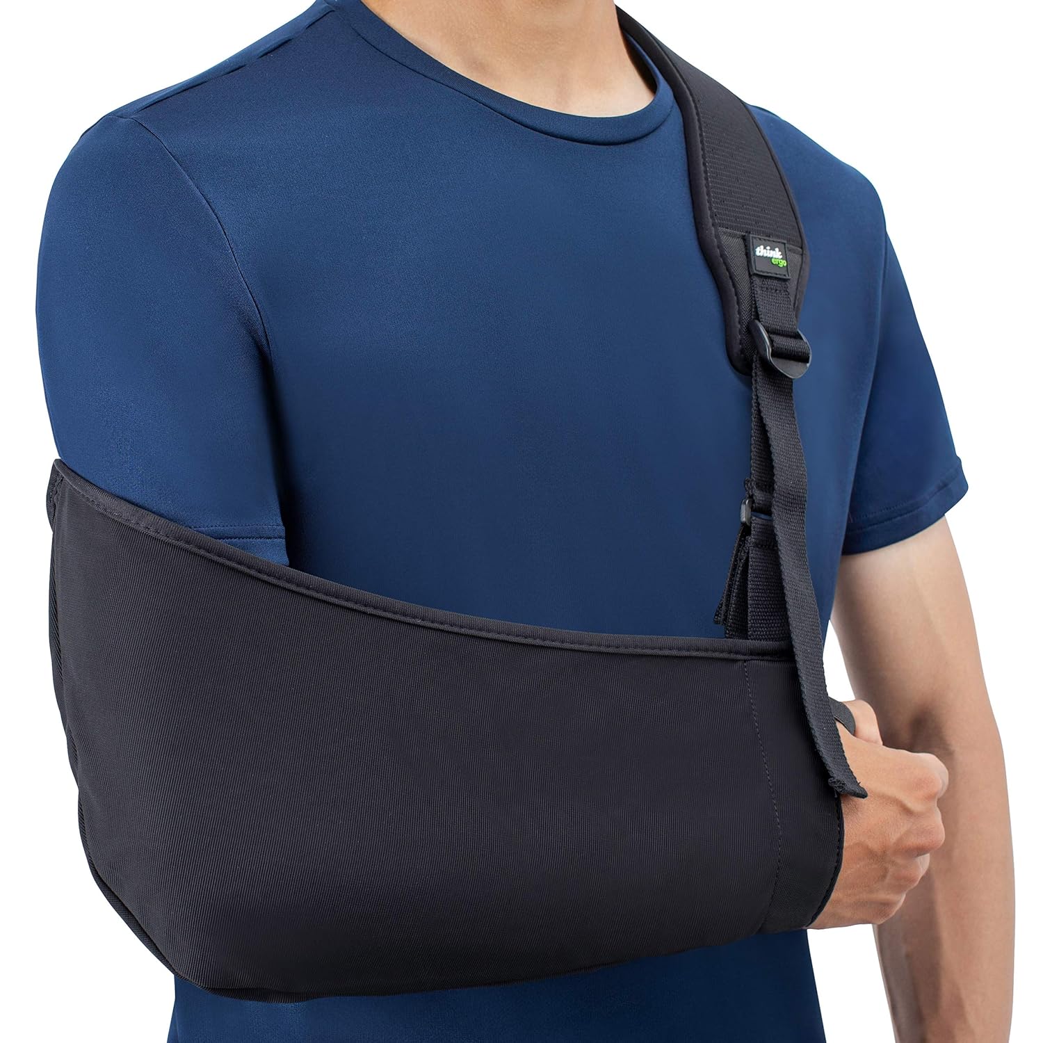
- Immobilization: Shoulder Sling
- Discontinue once asymptomatic
- Type I: Typically 1-3 weeks
- Type II: Longer, up to 4 weeks[8]
- Activity modification
- Type II injuries may require early rehabilitation program with passive and active shoulder ROM exercises
- Rotator cuff, scapular stabilization, and trunk strengthening exercises as pain resolves
- Heavy lifting and contact activities only once extremity is pain free and symmetric ROM is acheived
- Consider Corticosteroid Injection in refractory cases
Type III
- Management is controversial
- Not a lot of high level evidence to guide decision making
- General consensus is to advocate for initial nonoperative management.
- Individualized treatment based on patient activity level, impairment, and occupation
- Consider surgical repair in acute, young patients
- Comparably high satisfaction with operative and nonoperative treatment though higher complication rates in those treated surgically[9]
- Nonoperative treatment
- Similar to Type I, II
Type IV - VI
- Generally considered surgical
- Technique
- Many described in literature
- ORIF most common
Rehab and Return to Play
Rehabilitation
- Needs to be updated
Return to Play
- Return to play once pain completely resolved and equal active ROM in bilateral shoulders
- Followed by adequate strength training
- Recovery generally takes 6 weeks for Type II injuries and 12 weeks for Type III injuries
Prognosis and Complications
Prognosis
- Needs to be updated
Complications
- ChronicAcromioclavicular Joint Pain
- Experienced by 1/3 of patients at 6 months after injury and up to 6 years of follow up[10]
- Decrease in bench press strength (need citation)
- Cosmetic deformity is very common
- Crepitus, clicking
- AC Joint Arthritis
- Distal Clavicle Osteolysis
See Also
Internal
- Physical Exam Shoulder
- Shoulder Anatomy (Main)
- Neck Pain (Main)
- Back Pain (Main)
- Shoulder Pain (Main)
External
- Sports Medicine Review Shoulder Pain: https://www.sportsmedreview.com/by-joint/shoulder/
References
- ↑ Image courtesy of radiologymasterclass.co.uk
- ↑ . Tischer T, Salzmann GM, El-Azab H, Vogt S, Imhoff AB. Incidence of associated injuries with acute acromioclavicular joint dislocations types III through V. Am J Sports Med. 2009 Jan;;37(1):136-9. Epub 2008 Aug 25.
- ↑ Manske, Robert, and Todd Ellenbecker. "Current concepts in shoulder examination of the overhead athlete." International journal of sports physical therapy 8.5 (2013): 554.
- ↑ Kang, Ki-Ser, et al. "Long term follow up results of the operative treatment of the acromioclavicular joint dislocation with a Wolter plate." Journal of the Korean Fracture Society 22.4 (2009): 259-263.
- ↑ Manske, Robert C., et al. "MSK Diagnostic Ultrasound for the Assessment of the Acromioclavicular Joint." International Journal of Sports Physical Therapy 19.1 (2024): 1516.
- ↑ Zanca P. Shoulder pain: involvement of the acromioclavicular joint. (Analysis of 1,000 cases). Am J Roentgenol Radium Ther Nucl Med. 1971 Jul;112(3):493-506.
- ↑ Case courtesy of Andrew Murphy, Radiopaedia.org, rID: 72436
- ↑ Park JP, Arnold JA, Coker TP, Harris WD, Becker DA. Treatment of acromioclavicular separations. A retrospective study. Am J Sports Med. 1980 Jul-Aug;8(4):251-6
- ↑ Glick JM, Milburn LJ, Haggerty JF, Nishimoto D. Dislocated acromioclavicular joint: follow-up study of 35 unreduced acromioclavicular dislocations. Am J Sports Med. 1977 Nov-Dec;5(6):264-70.
- ↑ Mouhsine E, Garofalo R, Crevoisier X, Farron A. Grade I and II acromioclavicular dislocations: results of conservative treatment. J Shoulder Elbow Surg. 2003 NovDec;12(6):599-602.
Created by:
John Kiel on 4 July 2019 08:23:12
Authors:
Last edited:
18 April 2025 00:47:02
Categories: