Acromioclavicular Joint Pain
Other Names
- AC Joint OA
- Acromioclavicular Joint Osteoarthritis
- Degenerative Joint Disease of the Acromioclavicular Joint
- AC Joint Pain
- Acromioclavicular Joint Pain
- Acromioclavicular Pain
- AC Joint Injury
- Acromioclavicular Joint Syndrome
- AC Joint Arthralgia
- AC Joint Dysfunction
- Distal Clavicle Pain
- AC Joint Arthritis Pain
Background
- This page refers to atraumatic and/or chronic causes of pain to the Acromioclavicular Joint (AC Joint)
- Traumatic Acromioclavicular Joint Separation is discussed separately
History
- Hippocrates (460-377 BC) noted that AC joint dislocations were often misdiagnosed as glenohumeral injuries
- First detailed description of AC joint injury pathoanatomy is widely credited to Cadenat in 1917[1]
Epidemiology
- Affects approximately 45 per 100,000 person-years
- AC joint injuries accounting for 11% of all shoulder injuries[2]
- Tends to affect mild aged (20-49 years) males, with 82% of cases ocurring in men
- Sports-related injuries account for 53% of AC joint injuries
Introduction


General
- AC Joint Pain is a common cause of shoulder pain that can result from traumatic injurise or degenerative osteoarthriitis
- The AC joint connects the clavicle to the acromion and is critical for shoulder stability and motion
- The diagnosis is made clinically and supported by imaging findings
- Patients are initially treated conservatively but may require surgical intervention in refractory cases
Etiology: Primary Osteoarthritis
- Refers to age-related degeneration, typically affecting middle aged individuals
- Most common cause of AC joint pain[7]
- Intra-articular disc
- Functions like the meniscus of the knee
- Prone to fraying, tearing, and forming holes, macerated by defects in the chondral surface
- Inflammatory arthropathies:
- Septic arthritis
- Uncommon in AC joint but risk factors include trauma, recent surgery, IV drug abuse, immune compromised, and hematogenous seeding among many others[8]
Etiology: Post Traumatic
- Most common in young adult males who play contact sports (football, hockey) or cycling[9]
- Most commonly occuring with an axial load on an abducted arm[10]
- Many of thesee athletes will have sustained AC Joint Separation or Dislocation
- Distal clavicle osteolysis
- Related to repetitive microtrauma[11]
- Most commonly in weight lifters, less commonly basketball, swimming
- Joint instability
- Due to local elevation of contact stresses, dynamic loss of joint congruity, and alterations in range of motion[12]
Anatomy of the Acromioclavicular Joint
- General
- Stabilizers
- Static: Acromioclavicular Ligament, Coracoclavicular Ligaments
- Dynamic: Deltoid, Trapezius, Serratus Anterior
Associated Injuries[13]
- Rotator Cuff Disease
- Includes: Rotator Cuff Tear, Rotator Cuff Tendonitis
- Condition most frequently associated condition with AC joint disorders
- Coexisting rotator cuff disease occurs in approximately 79-98% of cases
- Subacromial Impingement
- Frequently co-exist with AC joint pain and can be difficult to distinguish clinically
- Glenoid Labral Tears
- Superior labral anterior-posterior (SLAP) lesions and other labral tears are common
- 45% of younger patients (under 50), 29% of older patients (oveer 50) have labral tears[13]
- Proximal Biceps Tendon Injuries
- Glenohumeral Osteoarthritis
- Present in 14% of patients with symptomatic AC degeneration[14]
Risk Factors
Traumatic Risk Factors[15]
- Direct fall onto the lateral shoulder (most common mechanism)
- Contact sports (e.g., football, hockey, rugby)
- High-energy trauma (motor vehicle collisions, cycling accidents)
- Prior AC joint sprain or separation (predisposes to chronic pain and instability)
Repetitive / Overuse Risk Factors[10]
- Repetitive overhead activity (throwing athletes, swimmers)
- Weightlifting (especially bench press, dips, overhead press)
- Occupational overhead labor (construction, painting, manual labor)
- Chronic microtrauma leading to distal clavicle osteolysis
Degenerative Risk Factors[16]
- Increasing age (AC joint osteoarthritis is common after age 40)
- Prior joint injury accelerating degenerative changes
- Chronic mechanical stress across the AC joint
- Joint space narrowing and osteophyte formation
Anatomic / Biomechanical Risk Factors[17]
- Scapular dyskinesis altering AC joint loading
- Clavicular malalignment or prior fracture
- Ligamentous laxity or instability of the AC joint
- Abnormal shoulder kinematics (poor rotator cuff or periscapular control)
Sport-Specific Risk Factors[18]
- Contact athletes (football, rugby, wrestling)
- Overhead athletes (baseball, tennis, volleyball)
- CrossFit and heavy resistance training athletes
- Repetitive axial loading across the shoulder
Post-Surgical / Iatrogenic Risk Factors[19]
- Prior distal clavicle resection (persistent instability or pain)
- Failed AC joint reconstruction
- Altered biomechanics following shoulder surgery
Systemic / Other Risk Factors[20]
- Inflammatory arthritis (e.g., rheumatoid arthritis, pseudogout)
- Infection (rare, septic AC joint)
- Osteolysis related to metabolic or endocrine factors
- Tobacco Use Disorder (associated with impaired healing and degeneration)
Differential Diagnosis
Differential Diagnosis Shoulder Pain
- Fractures
- Proximal Humerus Fracture
- Humeral Shaft Fracture
- Clavicle Fracture
- Scapula Fracture
- First Rib Fracture (traumatic or atraumatic)
- Floating Shoulder
- Dislocations & Separations
- Arthropathies
- Muscle & Tendon Injuries
- Rotator Cuff
- Bursopathies
- Ligament Injuries
- Neuropathies
- Other
- Pediatrics
- Coracoid Avulsion Fracture
- Humeral Head Epiphysiolysis (Little League Shoulder)
Clinical Features


History
- Can be difficult to distinguish from other causes of shoulder pain
- Often other pathology co-occurs including rotator cuff tears, labral injuries and biceps tendonitis
- Patient may report pain with passive and active range of motion
- Pain typically anterior shoulder but can but can be referred to anterolateral neck, anterolateral deltoid, and trapezius[10]
- In patients with traumatic history, they should be able to describe a prior injury and treatment
- Degenerativee causes are often more insidious with no clear eetiology
- Pain descriptions may include[7]
- Pain exacerbated by overhead activities and cross-body adduction
- Pain localized to the AC joint without radiation below the elbow
Physical: Physical Exam Shoulder
- Inspection: AC Joint may demonstrate swelling, deformity, or prominence[23]
- Observe for asymmetry, deformity, swelling, or prominence of the distal clavicle
- Tenderness to AC joint is sensitive, not specific[24]
- In isolated injuries, range of motion is usually preserveed
Special Tests
- Crossover Test: Examiner passively flexs, adducts arm across body
- Resisted AC Joint Extension Test: Flex, internally rotatoe shoulder and abduct against resistance
- OBriens Test: Shoulder flexed to 90, flexes further against resistance
- Crossover test most sensitive (77%), O'Briens Test is most specific (95%)[25]
- One Finger Test: Have patient point to most painful spot with 1 finger (AC joint = positive)
- Paxinos Test: Apply pressure at the acromion and clavicle
- Sensitivity 79%, specificity 50%[26]
- AC Joint Line Tenderness Test: palpate the AC joint directly
- Sensitivity 96%, specificity 10%[26]
- Bell Van Riet Test: cross body test with resisted abduction
Evaluation


Radiographs
- Standard Radiographs Shoulder
- Initial imaging study of choice
- OA Findings: joint space narrowing, subchondral cysts, osteophytes, and subchondral sclerosis
- May see distal clavicle osteolysis
- Asymptomatic AC joint OA findings are common (need citation)
- Zanca View: 10-15° cephalid tilt best visualizes joint
- Weighted vs. Non-Weighted Views
CT
- Provides superior osseous visualization[30]
- Indications[31]
- Evaluating complex fracture patterns involving the distal clavicle
- Assessing bone quality before surgical reconstruction
- Detecting subtle osteophytes and subchondral changes
MRI
- General[32]
- MRI enables visualization of the capsuloligamentous structures
- Including the integrity of the AC joint ligaments, coracoclavicular ligaments
- Assocaited rotator cuff tears, labral pathology
- MRI Findings in Degenerative Disease[7]
- Bone marrow edema (observed only in symptomatic patients)
- Inferior joint distension
- Impression on the supraspinatus muscle
Ultrasound
- Diagnostic accuracy
- Advantages[33]
- Non-invasive, accessible, and cost-effective
- Can assess dynamic horizontal instability in real-time
- Useful for excluding joint inflammation
- Improves accuracy of diagnostic and therapeutic injections
- Diagnostic injection can help clarify etiology of shoulder pain
Classification
Acromioclavicular joint arthrosis classified by Shubin–Stein
- Grade I
- No capsular distension
- No joint space narrowing
- No evidence of osteophyte formation
- Grade II
- Capsular distension
- Occasional mild joint space narrowing
- Grade III
- Capsular distension
- Joint space narrowing
- Subacromial fat effacement
- Marginal osteophyte formation
- Grade IV
- Everything in grade I, II and III
- Marked joint space irregularity and narrowing with large osteophytes
Management
Nonoperative
- First line therapy in most patients
- Activity Modification
- Avoid: aggravating activities such as cross-body, pushing, weight lifting, throwing, overhead work
- Physical Therapy
- Can help with range of motion, flexibility, and strength[37]
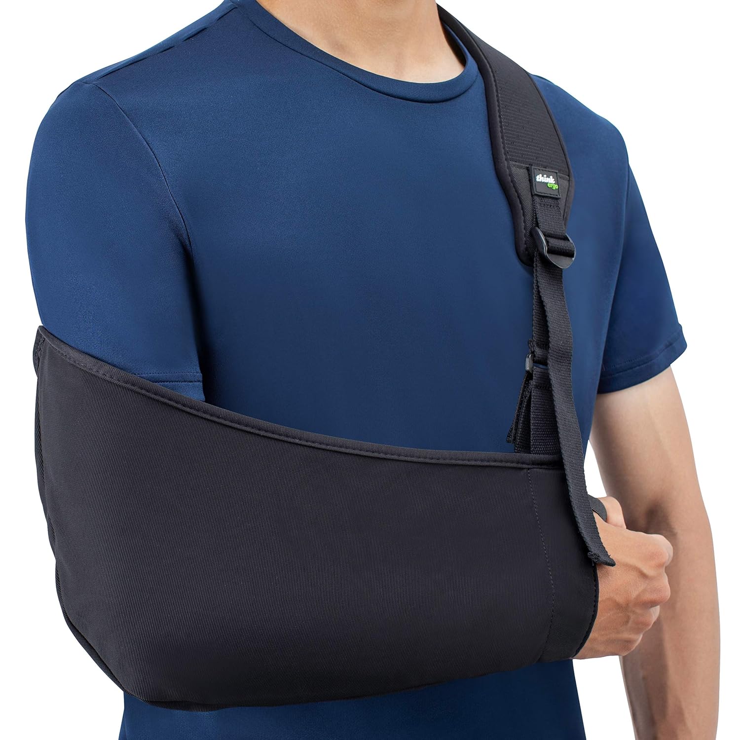
- Immobilization
- Temporary immobilization with Shoulder Sling in the setting of acute exacerbation
- Medications including NSAIDS, Acetaminophen
- Acromioclavicular Joint Injection
Operative
- Indications
- Failure of non-operative therapy, typically a minimum of 6 months
- Technique
- Arthroscopic clavicle resection
- Open clavicle resection (Mumford procedure)
Rehab and Return to Play


Rehabilitation
- General rehab objectives
- Pain control and restoration of range of motion (ROM)
- Scapular stabilization and rotator cuff strengthening
- Sport-specific conditioning and gradual return to activity
- Dictated in part by concomitant procedures[7]
- Distal clavicle excision
- Immobilized in shoulder sling, allowed to perform pendulums
- 2 weeks: begin passive, active range of motion
- 4 weeks: discontinue immobilization, full range of motion permitted
- Note: If significant deltoid dissection, flexion and abduction restricted up to 6 weeks
Phase 1: Acute/Protection Phase (Weeks 0-2)
- Goals: Pain and inflammation control, tissue protection, maintain elbow/wrist/hand mobility
- Immobilization
- Broad-arm sling for comfort (typically 2-4 weeks depending on injury severity)
- Remove sling 4 times daily for gentle exercises
- Pain Management
- Ice application: 15-20 minutes every 2-3 hours
- Oral NSAIDs or analgesics as prescribed
- Activity modification: Avoid overhead activities, cross-body movements, and heavy lifting
- Exercises
- Pendulum exercises (Codman's): Gentle circular motions using gravity, 2-3 minutes, 4x daily
- Elbow, wrist, and hand range of motion exercises
- Cervical spine gentle range of motion
- Scapular squeezes (gentle isometric retraction in neutral position)
- Criteria to Progress: Pain at rest 3/10, able to tolerate passive range of motion
Phase 2: Early Mobility Phase (Weeks 2-4)
- Goals: Restore passive and active-assisted range of motion, begin gentle strengthening
- Range of Motion:
- Passive and active-assisted forward flexion (supine, using opposite arm or wand)
- Passive external rotation with arm at side
- Active-assisted abduction in scapular plane
- Avoid cross-body adduction and end-range overhead positions
- Early Strengthening:
- Isometric exercises in neutral position:
- Shoulder flexion, extension, abduction (submaximal, pain-free)
- External and internal rotation isometrics
- Scapular clock exercises (gentle protraction/retraction)
- Scapular Stabilization (Early Stage):
- Low row exercise (activates serratus anterior and lower trapezius at moderate levels)
- Inferior glide exercise
- Wall slides (limited range)
- Frequency: 2-3 sets of 10-15 repetitions, 2x daily
- Criteria to Progress: Near-full passive range of motion, minimal pain with active motion
Phase 3: Intermediate Strengthening Phase (Weeks 4-8)
- Goals: Restore full active range of motion, progressive strengthening, improve scapular control
- Active Range of Motion:
- Full active forward flexion and abduction
- External rotation at 0° and progressing to 45° abduction
- Begin gentle cross-body stretching if tolerated
- Strengthening (Resistance Bands/Light Weights):
- External rotation in side-lying position (excellent scapular muscle balance)
- Internal rotation with resistance band
- Prone horizontal abduction with external rotation (optimal scapular neuromuscular control)
- Prone extension
- Biceps curls, triceps extensions
- Scapular Stabilization (Progressive):
- Lawnmower exercise (activates serratus anterior and lower trapezius)
- Robbery exercise
- Wall push-up plus (serratus anterior activation)
- Scapular retraction with resistance band
- Prone Y, T, W exercises
- Frequency: 3 sets of 10-15 repetitions, daily
- Criteria to Progress: Full pain-free active range of motion, 4/5 strength in all planes
Phase 4: Advanced Strengthening Phase (Weeks 8-12)
- Goals: Restore full strength, begin sport/activity-specific training
- Progressive Resistance Training:
- Shoulder press (avoid extreme overhead positions initially)
- Rows (seated, bent-over)
- Lat pulldowns
- Push-ups (floor progression)
- Dumbbell exercises in functional patterns
- Scapular Stabilization (Advanced):
- Push-up plus on unstable surface
- Quadruped exercises with arm lifts
- Plank variations with shoulder taps
- Serratus punches
- Proprioception and Neuromuscular Control:
- Rhythmic stabilization exercises
- Ball tosses against wall
- Perturbation training
- Closed kinetic chain exercises (weight bearing through upper extremity)
- Frequency: 3-4 sets of 8-12 repetitions, 3-4x weekly
- Criteria to Progress: Strength equal to contralateral side, no pain with resistance exercises
AC Joint Pain Rehab Exercises PDF
- AC Joint Injury Rehabilitation PDF
- AC Joint Sprain Rehab Exercises PDF
- Mild acromioclavicular AC Joint Injury Rehab PDF
- Osteoarthritis of the Acromioclavicular Joint PDF
Return to Play

- Goals: Full return to work, sport, and recreational activities
- Criteria for Return to Activity:
- Normal shoulder motion compared to contralateral side
- Strength equal to contralateral extremity (≥90%)
- Shoulder is asymptomatic with sport-specific movements
- No pain with provocative maneuvers (cross-body adduction)
- Successful completion of sport-specific drills
- Sport-Specific Progression:
- Gradual return to sport-specific movements
- Contact sports: Progress from non-contact drills to controlled contact to full participation
- Overhead athletes: Interval throwing/serving programs
- Consider protective padding for contact sports during initial return
- Average time lost
- Low-grade, atraumatic conditions, the average time lost to sport is 10–18 days[39]
- Most athletes experience significant improvement with appropriate treatment within 4-6 weeks
Prognosis and Complications

Prognosis
- General
- Most patients have favorable long term prognosis with appropriate treatment
- High likelihood of return to previous activity levels and minimal long-term disability
- Return to baseline function is expected in weeks to a few months for mild cases.[40]
- Surgical outcomes
- Distal clavicle excision provides high rates of relief and patient satisfcation[7]
Complications
- Chronic pain
- Inability to return to sport
- AC Joint Osteoarthritis
- Subacromial Impingement Syndrome
- Rotator Cuff Tendinopathy
- Scapular Dyskinesis
- Distal Clavicle Osteolysis
- Surgical
- AC Joint instability due to excessive clavicle resection
- Persistent pain due to incomplete resection
- Heterotopic Ossification
- Deltoid dehiscence
See Also
Internal
External
- Sports Medicine Review Shoulder Pain: https://www.sportsmedreview.com/by-joint/shoulder/
References
- ↑ Cadenat, F. M. "Treatment of fractures and dislocations, outer end, clavicle." Internat. Clin. 27 (1917): 145-169.
- ↑ Skjaker, Stein Arve, et al. "Young men in sports are at highest risk of acromioclavicular joint injuries: a prospective cohort study." Knee surgery, sports traumatology, arthroscopy 29.7 (2021): 2039-2045.
- ↑ Image courtesy of teachmeanatomy, "The Acromioclavicular Joint"
- ↑ Image courtesy of radiologymasterclass.co.uk
- ↑ Chalmers, Peter N., et al. "Preoperative factors associated with subsequent distal clavicle resection after rotator cuff repair." Orthopaedic Journal of Sports Medicine 7.5 (2019): 2325967119844295.
- ↑ Thomas, Jija, Makki Daud, and Simon Macmull. "Acute septic arthritis of the acromioclavicular joint caused by Staphylococcus aureus with marked soft tissue collection towards posterior medial aspect of the AC joint: A rare clinical presentation." IDCases 29 (2022): e01513.
- ↑ 7.0 7.1 7.2 7.3 7.4 Mall, Nathan A., et al. "Degenerative joint disease of the acromioclavicular joint: a review." The American journal of sports medicine 41.11 (2013): 2684-2692.
- ↑ Bossert, M, Prati, C, Bertolini, E, Toussirot, E, Wendling, D. Septic arthritis of the acromioclavicular joint. Joint Bone Spine. 2010;77(5):466-469.
- ↑ Flores, Dyan V., et al. "Imaging of the acromioclavicular joint: anatomy, function, pathologic features, and treatment." Radiographics 40.5 (2020): 1355-1382.
- ↑ 10.0 10.1 10.2 Mazzocca, Augustus D., et al. “Acromioclavicular Joint Injuries: Diagnosis and Management.” Journal of the American Academy of Orthopaedic Surgeons, vol. 15, no. 5, 2007, pp. 267–278.
- ↑ Charron, KM, Schepsis, AA, Voloshin, I. Arthroscopic distal clavicle resection in athletes: a prospective comparison of the direct and indirect approach. Am J Sports Med. 2007;35(1):53-58.
- ↑ Shu, B, Johnston, T, Lindsey, DP, McAdams, TR. Biomechanical evaluation of a novel reverse coracoacromial ligament reconstruction for acromioclavicular joint separation. Am J Sports Med. 2012;40(2):440-446.
- ↑ 13.0 13.1 Brown, JN, Roberts, SN, Hayes, MG, Sales, AD. Shoulder pathology associated with symptomatic acromioclavicular joint degeneration. J Shoulder Elbow Surg. 2000;9(3):173-176.
- ↑ Markel, Jochen, et al. "Concomitant glenohumeral pathologies in high-grade acromioclavicular separation (type III–V)." BMC Musculoskeletal Disorders 18.1 (2017): 439.
- ↑ Beitzel, Knut, et al. “Current Concepts in the Treatment of Acromioclavicular Joint Dislocations.” Arthroscopy, vol. 29, no. 2, 2013, pp. 387–397.
- ↑ Cadogan, Alex, et al. “A Prospective Study of Shoulder Pain in Primary Care: Prevalence of Imaged Pathology and Response to Guided Diagnostic Blocks.” BMC Musculoskeletal Disorders, vol. 12, 2011, p. 119.
- ↑ Kibler, W. Ben, et al. “Scapular Dyskinesis and Its Relation to Shoulder Pain.” Journal of the American Academy of Orthopaedic Surgeons, vol. 20, no. 6, 2012, pp. 364–372.
- ↑ Trainer, George, et al. “Distal Clavicle Osteolysis in Weight Lifters.” The American Journal of Sports Medicine, vol. 14, no. 4, 1986, pp. 295–298.
- ↑ Cook, J. B., et al. “Acromioclavicular Joint Injuries: Evidence-Based Treatment.” Journal of the American Academy of Orthopaedic Surgeons, vol. 26, no. 7, 2018, pp. e136–e146.
- ↑ Rockwood, Charles A., et al. The Shoulder. 5th ed., Elsevier, 2016.
- ↑ Image courtesy of https://musculoskeletalkey.com/, "Disorders of the Acromioclavicular Joint"
- ↑ Almoallim, Hani, et al. "Approach to Musculoskeletal Examination." Skills in Rheumatology (2021): 17-65.
- ↑ Chronopoulos, E, Kim, TK, Park, HB, Ashenbrenner, D, McFarland, EG. Diagnostic value of physical tests for isolated chronic acromioclavicular lesions. Am J Sports Med. 2004;32(3):655-661.
- ↑ Hegedus, EJ, Goode, A, Campbell, S. Physical examination tests of the shoulder: a systematic review with meta-analysis of individual tests. Br J Sports Med. 2008;42(2):80-92; discussion 92.
- ↑ Chronopoulos, E, Kim, TK, Park, HB, Ashenbrenner, D, McFarland, EG. Diagnostic value of physical tests for isolated chronic acromioclavicular lesions. Am J Sports Med. 2004;32(3):655-661.
- ↑ 26.0 26.1 Walton, Judie, et al. "Diagnostic values of tests for acromioclavicular joint pain." JBJS 86.4 (2004): 807-812.
- ↑ Eriks-Hoogland, I., et al. "Acromioclavicular joint arthrosis in persons with spinal cord injury and able-bodied persons." Spinal cord 51.1 (2013): 59-63.
- ↑ Ibrahim, E. F., N. P. Forrest, and A. Forester. "Bilateral weighted radiographs are required for accurate classification of acromioclavicular separation: an observational study of 59 cases." Injury 46.10 (2015): 1900-1905.
- ↑ Nordin, Jonas S., et al. "Weighted or internal rotation radiographs are not useful in the classification of acromioclavicular joint dislocations." Acta Radiologica 62.6 (2021): 758-765.
- ↑ Ernberg, LA, Potter, HG. Radiographic evaluation of the acromioclavicular and sternoclavicular joints. Clin Sports Med. 2003;22(2):255-275.
- ↑ 31.0 31.1 Alasaarela, E., et al. "Ultrasound evaluation of the acromioclavicular joint." The Journal of rheumatology 24.10 (1997): 1959-1963.
- ↑ Flores, Dyan V., et al. "Imaging of the acromioclavicular joint: anatomy, function, pathologic features, and treatment." Radiographics 40.5 (2020): 1355-1382.
- ↑ 33.0 33.1 Faruch Bilfeld, Marie, et al. "Ultrasound of the coracoclavicular ligaments in the acute phase of an acromioclavicular disjonction: Comparison of radiographic, ultrasound and MRI findings." European radiology 27.2 (2017): 483-490.
- ↑ Tat, Jimmy, Jessica Tat, and John Theodoropoulos. "Clinical applications of ultrasonography in the shoulder for the orthopedic surgeon: a systematic review." Orthopaedics & Traumatology: Surgery & Research 106.6 (2020): 1141-1151.
- ↑ Borbas, Paul, et al. "The influence of ultrasound guidance in the rate of success of acromioclavicular joint injection: an experimental study on human cadavers." Journal of shoulder and elbow surgery 21.12 (2012): 1694-1697.
- ↑ Sabeti-Aschraf, Manuel, et al. "Ultrasound guidance improves the accuracy of the acromioclavicular joint infiltration: a prospective randomized study." Knee Surgery, Sports Traumatology, Arthroscopy 19.2 (2011): 292-295.
- ↑ Docimo, S, Kornitsky, D, Futterman, B, Elkowitz, DE. Surgical treatment for acromioclavicular joint osteoarthritis: patient selection, surgical options, complications, and outcome. Curr Rev Musculoskelet Med. 2008;1(2):154-160.
- ↑ 38.0 38.1 38.2 LeVasseur, Matthew R., et al. "Acromioclavicular joint injuries: effective rehabilitation." Open access journal of sports medicine (2021): 73-85.
- ↑ Pallis, Mark, et al. "Epidemiology of acromioclavicular joint injury in young athletes." The American journal of sports medicine 40.9 (2012): 2072-2077.
- ↑ Beitzel, K., et al. “Current Concepts in the Treatment of Acromioclavicular Joint Dislocations.” Arthroscopy, vol. 29, no. 2, 2013, pp. 387–397.