Coracoclavicular Bursa
Description
-
-
-
![(A, B) Sagittal PD FS sequence images at different plane levels demonstrate coracoclavicular bursal fluid distension (arrow) with narrowing of the coracoclavicular distance (2 mm)[1]](/w/images/thumb/9/94/MRI_coracoclavicular_bursitis.jpeg/120px-MRI_coracoclavicular_bursitis.jpeg) (A, B) Sagittal PD FS sequence images at different plane levels demonstrate coracoclavicular bursal fluid distension (arrow) with narrowing of the coracoclavicular distance (2 mm)[1]
(A, B) Sagittal PD FS sequence images at different plane levels demonstrate coracoclavicular bursal fluid distension (arrow) with narrowing of the coracoclavicular distance (2 mm)[1] -
![Sagittal PD FS sequence images at different plane levels demonstrate coracoclavicular bursal fluid distension (arrow) with narrowing of the coracoclavicular distance (2.2 mm)[1]](/w/images/thumb/7/77/Coracoclavicular_bursitis_MRI.jpeg/120px-Coracoclavicular_bursitis_MRI.jpeg) Sagittal PD FS sequence images at different plane levels demonstrate coracoclavicular bursal fluid distension (arrow) with narrowing of the coracoclavicular distance (2.2 mm)[1]
Sagittal PD FS sequence images at different plane levels demonstrate coracoclavicular bursal fluid distension (arrow) with narrowing of the coracoclavicular distance (2.2 mm)[1] -
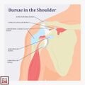
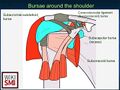
 Bursa aorund the shoulder joint: 1. subacromial-subdeltoid bursa 2. Subscapularis bursa 3. Sucoracoid bursa 4. Coracoclavicular bursa 5. Supra-acromial bursa.


![(A, B) Sagittal PD FS sequence images at different plane levels demonstrate coracoclavicular bursal fluid distension (arrow) with narrowing of the coracoclavicular distance (2 mm)[1]](/File:MRI_coracoclavicular_bursitis.jpeg)
![Sagittal PD FS sequence images at different plane levels demonstrate coracoclavicular bursal fluid distension (arrow) with narrowing of the coracoclavicular distance (2.2 mm)[1]](/File:Coracoclavicular_bursitis_MRI.jpeg)


Name
- Coracoclavicular Bursa
- Supracoracoid bursa
- Coracoclavicular ligament bursa
- Conoid–trapezoid bursa
- Bursa of the coracoclavicular ligaments
- Subcoracoid–coracoclavicular bursa
- Coracoid–clavicular bursa
General
- Found within the angle separating the trapezoid/conoid segments of the coracoclavicular ligament[4]
- Surrounded by various amounts of fibro fatty tissue
- Smaller discrete bursae around the coracoid process can be found adjacently
- One of the minor bursa of the shoulder
Anatomic Variance
- Bursa may be absent and the trapezoid/conoid ligaments are separated by adipose tissue
Function
- Permits frictionless movement between the coracoclavicular ligament and the clavicle
Clinical Significance
- Calcific coracoclavicular bursitis
- Septic Bursitis
See Also
References
- ↑ 1.0 1.1 Obaid, Haron, et al. "Coracoclavicular bursal changes on MRI: a diagnostic consideration in patients with shoulder pain and reduced coracoclavicular distance." Skeletal Radiology 51.9 (2022): 1837-1841.
- ↑ Image courtesy of elsevier.com
- ↑ Case courtesy of Henry Knipe, Radiopaedia.org, rID: 31397
- ↑ Bureau, N. J., Robert G. Dussault, and Theodore E. Keats. "Imaging of bursae around the shoulder joint." Skeletal radiology 25 (1996): 513-517.