Metatarsophalangeal Joint
(Redirected from First metatarsophalangeal joint)
Introduction
-
-
-

-
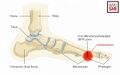
![The anatomy of the plantar aspect of the 1st MTP joint is illustrated and labeled[1]](/w/images/thumb/5/53/1st_MTPJ_plantar.jpg/120px-1st_MTPJ_plantar.jpg) The anatomy of the plantar aspect of the 1st MTP joint is illustrated and labeled[1]
The anatomy of the plantar aspect of the 1st MTP joint is illustrated and labeled[1] -
-


![The anatomy of the plantar aspect of the 1st MTP joint is illustrated and labeled[1]](/File:1st_MTPJ_plantar.jpg)


Names
- MTP joints
- Toe joints
- Metatarsal-phalangeal joints
- Forefoot joints
- Ball of foot joints
- Metatarsophalangeal articulations
- Metatarsal head joints
- Phalangeal base joints
- First MTP joint (for great toe specifically)
- Digital joints of the foot
General
- Synovial joints located between the heads of the metatarsal bones and the bases of the proximal phalanges of the toes[8]
- Each joint is stabilized by a fibrous capsule, collateral ligaments, and a plantar plate
- The first MTP joint has additional sesamoid bones embedded in its plantar aspect
Function
- Joints allow flexion, extension, and limited abduction/adduction
- Contribute to the push-off phase of gait and overall forefoot stability
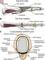
1st Metatarsophalangeal Joint
General
- Hinged ball and socket articulation
- Formed between the first metatarsal and its corresponding proximal phalanx[9]
- Because of the shallow concavity of the base of the proximal phalanx, this articulation is relatively unstable
- Stabilized by 9 ligaments, 3 tendons and 2 sesamoid bones[10]
Tendon Attachments
- Flexor Hallucis Brevis has two slips, which attach to and contain the medial (tibial) and lateral (fibular) sesamoid, respectively
- Abductor Hallucis attaches to medial sesamoid
- Adductor Hallucis attaches to lateral sesamoid
- Articulate with a facet on the plantar surface of the first metatarsal head
- Add stability to the joint
- Protects the tendons in which they are housed
- Increase the mechanical advantage of the FHB to facilitate load distribution, act as a shock absorber
- Normal range of motion displaces the sesamoids relative to the metatarsal head
Ligaments[12]
- 2 collateral ligaments which stabilize the joint during cutting
- 2 plantar ligaments
- Sometimes an intraarticular meniscus that further enhances stability
- Formed by the plantar ligaments, thickened plantar capsule and tendons of the FHB
- Orignates from the head of the metatarsal at the distal portion of the sesamoids
- Inserts to the plantar base of the proximal phalanx.
Range of Motion
- Dorsiflexion: approximately 50° of dorsiflexion
- Plantarflexion: 30° of plantarflexion
Weight Bearing[14]
- Normal gait: sustain 40% to 60% of body weight
- Walking: 80% of body weight during the toe-off phase of normal
- This force increases 2x to 8x body weight when running and jumping[15]
Vascular Supply
- First dorsal metatarsal artery
- First plantar metatarsal artery
- Branches of the medial plantar artery
Innervation
2nd to 5th MTP Joints
General
- Condyloid joints
- Rounded head of the metatarsal articulates with the shallow cavity of the proximal phalanx
- Enclosed by a fibrous capsule
- Reinforces by collateral ligaments and a plantar plate
Intrinsic Muscles
- Flexor Digiti Minimi Brevis
- Lumbricals of the Foot
- Plantar Interossei of the Foot
- Dorsal Interossei of the Foot
- Flexor Digitorum Brevis
- Abductor Digiti Minimi of the Foot
- Extensor Digitorum Brevis
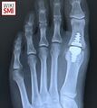
Clinical Significance
1st MTPJ
See Also
References
- ↑ Case courtesy of Matt Skalski, Radiopaedia.org, rID: 45171
- ↑ Nery, Caio, et al. "First MTP joint instability—expanding the concept of “turf-toe” injuries." Foot and Ankle Surgery 26.1 (2020): 47-53.
- ↑ Nery, Caio, Hilary Umans, and Daniel Baumfeld. "Etiology, clinical assessment, and surgical repair of plantar plate tears." Seminars in musculoskeletal radiology. Vol. 20. No. 02. Thieme Medical Publishers, 2016.
- ↑ Image courtesy of www.imaios.com/en
- ↑ Light, Jonathan, Laurie L. Wellman, and Richard M. Conran. "Educational Case: Gout." Academic Pathology 10.1 (2023): 100065.
- ↑ Sherman, Thomas I., et al. "First metatarsophalangeal joint arthroscopy for osteochondral lesions." Arthroscopy Techniques 5.3 (2016): e513-e518.
- ↑ Yamauchi, Junichiro, and Keiji Koyama. "The mechanical role of the metatarsophalangeal joint in human jumping." Plos one 17.5 (2022): e0268634.
- ↑ Drago, Sebastián, et al. "Assessment and management of atraumatic first metatarsophalangeal joint pain." JAAOS-Journal of the American Academy of Orthopaedic Surgeons 31.14 (2023): 708-716.
- ↑ York PJ, Wydra FB, Hunt KJ. Injuries to the great toe. Curr Rev Musculoskelet Med. 2017;10:104-112. doi:10.1007/s12178-017- 9390-y
- ↑ Hong CC, Pearce CJ, Ballal MS, Calder JD. Management of sports injuries of the foot and ankle: an update. Bone Joint J. 2016;98-B:1299-1311. doi:10.1302/0301- 620X.98B10.37896
- ↑ Kadakia AR, Molloy A. Current concepts review: traumatic disorders of the first metatarsophalangeal joint and sesamoid complex. Foot Ankle Int. 2011;32:834-839. doi:10.3113/FAI.2011.0834
- ↑ Childs SG. The pathogenesis and biomechanics of turf toe. Orthop Nurs. 2006;25:276-280. doi:10.1097/00006416- 200607000-00012
- ↑ Hong CC, Pearce CJ, Ballal MS, Calder JD. Management of sports injuries of the foot and ankle: an update. Bone Joint J. 2016;98-B:1299-1311. doi:10.1302/0301- 620X.98B10.37896
- ↑ Stokes IA, Hutton WC, Stott JR, Lowe LW. Forces under the hallux valgus foot before and after surgery. Clin Orthop Relat Res. 1979;(142):64-72.
- ↑ Nigg BM, Yeadon MR. Biomechanical aspects of playing surfaces. J Sports Sci. 1987;5:117-145. doi:10.1080/02640418708729771