Retrocalcaneal Bursitis
Other Names
- Subcutaneus Calcaneal Bursitis
- Albert disease
- Calcaneus altus
- Pump bump
- Winter heel
- Achillodynia
- Chronic retrocalcaneal bursitis
- Subtendinous Bursitis
- Achilles bursitis
- Posterior heel bursitis
- Retro-Achilles bursitis
- Calcaneal bursitis
- Bursitis of the retrocalcaneal bursa
Background
- This page refers to inflammation or bursitis of the Retrocalcaneal Bursa
- Note that Haglunds Deformity is a discrete disease process that can present similarly
History
- First described by Painter in 1898[1]
Epidemiology
- The epidemiology of retroachilles bursitis is poorly described in the literature
Pathophysiology


General
- Inflammation of the bursa between the Achilles tendon and the posterior calcaneus
- Typically results as a result of repeated microtrauma to the bursa
- Patients present with erythema and swelling of the region, tenderness to palpation
- Diagnosis is clinical and confirmed with imaging
- Management is nonsurgical including activity modification, NSAIDS and a walking boot
Etiology
- Repetitive impingement of the bursa between the anterior aspect of the Achilles tendon and a bony posterosuperior calcaneal prominence[5]
- This chronic microtrauma leads to inflammation of the bursa
Mechanism of Injury
- General
- Compression and chafing of the retrocalcaneal bursa during repetitive ankle motion
- During dorsiflexion, the bursa becomes compressed between anterior surface of the Achilles tendon, posterior calcaneal tuberosity[6]
- This repeated impingement causes inflammatory changes
- Anatomic Factors[7]
- Flat surface of the calcaneal tuberosity increases risk fourfold
- Calcaneal slope above 25° increases risk 2.8-fold
- Presence of Haglund deformity creates additional mechanical irritation
- Increased Achilles tendon thickness and smaller bursal surface area also contribute to impingement by further narrowing the retrocalcaneal space
Associated Conditions
Anatomy of the Retrocalcaneal Bursa
- Lies between the Calcaneus anteriorly and the Achilles Tendon posteriorly
- Separated from Achilles fat pad by synovial lining on superior aspect[8]
- Anterior wall is cartilaginous, posterior wall is tendinous
Risk Factors
- Sports
- Biomechanical
- Hindfoot Varus
- Rigid plantarflexed first ray
- Systemic
Differential Diagnosis
Differential Diagnosis Ankle Pain
- Fractures & Dislocations
- Muscle and Tendon Injuries
- Ligament Injuries
- Bursopathies
- Nerve Injuries
- Arthropathies
- Pediatrics
- Fifth Metatarsal Apophysitis (Iselin's Disease)
- Calcaneal Apophysitis (Sever's Disease)
- Triplane Fracture
- Other
Clinical Features

History
- Patients typically report posterior heel pain
- Worse with pressure from shoes
- Relieved when walking barefoot
- Swelling is often but not always present
- Typically worse during early exercise and improves during the workout
- Patients may report wearing ill fitting footwear
Physical Exam: Physical Exam Ankle
- Swelling, erythema at the bursa and posteiror calcaneus may be noted (i.e. 'pump bump')
- If haglund deformity present, there is a bony prominence of the superior aspect of the posterior calcaneus
- Tenderness to direct palpation of the posterior heel
- Passive dorsiflexion often induces pain
- Active plantarflexion reproduces pain
Special Tests
- Two Finger Squeeze Test: Pressure is applied with the fingers placed medially and laterally anterior to the Achilles tendon insertion
Evaluation


Radiographs
- Standard Radiographs Ankle
- Typically normal
- May show loss of retrocalcaneal recess
- Obliteration of the retrocalcaneal recess
- radiolucent area at the posteroinferior corner of Kager's triangle
- Sensitive at 79-83%, highly specific 98-100%[11]
- Additional findings[13]
- Prominent posterosuperior calcaneal tuberosity
- Thickening of the distal Achilles tendon outline
Ultrasound
- Can easily detect the retrocalcaneal bursa and fluid collection
- Only 50% sensitivity compared to MRI for identifying retrocalcaneal bursitis[14]
- Patient positioning
- Best performed with the patient laying prone
- Findings
- Triangular hypoechoic lesion situated between the Achilles tendon and the calcaneus
- Power doppler: increased blood flow around an abnormal bursa
- Advantages[15]
- Allows easy evaluation, comparison of both ankles
- More rapid and cost-effective than MRI
- Allows dynamic assessment
- Guide interventional procedures
- Perform serial follow up examinations
MRI
- General
- Not required to make diagnosis
- Most comprehensive and accurate imaging modality
- Normal bursa[16]
- May contain detectable fluid (average 1 mm anteroposterior, 6 mm transverse, 3 mm craniocaudal)
- Dimensions exceeding 1 mm anteroposteriorly, 11 mm transversely, or 7 mm craniocaudally indicate pathology
- Findings[13]
- Bursa will appear as an enlarged, fluid-filled structure
- Low signal intensity on T1-weighted images
- High signal intensity on fluid-sensitive images
- Other potential findings[17]
- Increased signal intensity and thickening of the Achilles tendon
- Prominence of the posterior calcaneus tuberosity
- Reactive marrow edema.
Classification
- Not applicable
Management

Nonoperative
- Indications
- Virtually all cases
- Ice Therapy
- Activity modification
- NSAIDS
- Physical Therapy
- Emphasis on stretching the Achilles tendon
- Microcurrent therapy
- One study suggested this was helpful as an adjunct when combined with standard therapy[19]
Medical Equipment and DME
- Footwear modification
- Shoes that are open backed may relieve pressure or tension
- Walking barefoot can also provide relief
- Heel Cup
- Raises the heel, offloading the bursa and achilles tendon
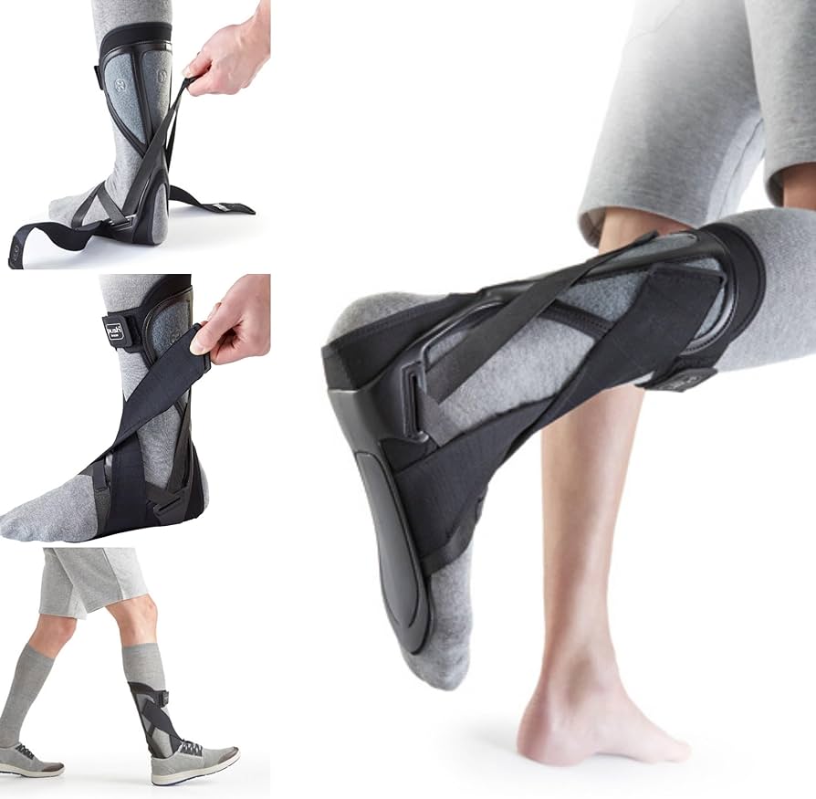
- Ankle Foot Orthosis
- When combined with stretching program, showed 88% success rate[20]
Procedural
- Corticosteroid Injection
- Performed under ultrasound or fluoroscopic guidance
- There is likely some risk to the Achilles Tendon, although this is not clearly established[21][22]
- Under fluoroscopy, patients reported a 50% reduction in pain[23]
- Risk of Achilles tendon rupture following injection is approximately 1.8%, typically occurring 15-59 days post-injection in association with acute injury[24]
Operative
- Indications
- Refractory to conservative management
- Technique
- Bursectomy
- Calcaneal resection
Rehab and Return to Play
Rehabilitation
- Structured stretching program
- Has the strongest evidence for success for achieving nonoperative success
- Especially when combined with an AFO
- Home stretching vs physical therapy[25]
- Both approaches produced similar outcomes at 6 weeks and 1 year
- Eccentric exercise program
- For associated Achilles tendinopathy, eccentric training improves VISA-A scores from 60.7 to 89.4 at 1 year
Rehab Program PDFs
Return to Play/ Work
- RTS continuum requires[26]
- Restoration of pain-free function and sport-specific performance
- Individualized assessment considering the athlete's sport, position, and timing in season
- Ongoing communication between the team physician, athlete, certified athletic trainers, and rehabilitation team
- Equipment modifications or bracing as necessary
Prognosis and Complications

Prognosis
- Conservative management
- There is an 88% success rate in avoiding surgery using structured nonoperative protocols
- Approximately 11-14% of patients progress to surgical intervention despite conservative treatment[24]
- Short-term outcomes with conservative treatment
- Significant improvement, with mean Foot Function Index scores improving from 48.4 to 18.6
- Image-guided corticosteroid injections yield excellent or good short-term response in 63-69% of patients
- Long-term surgical outcomes[28]
- Excellent when conservative measures fail
- Endoscopic calcaneoplasty demonstrates high patient satisfaction, good functional outcomes at a follow-up of 101 months
- Traditional open surgical approaches also show favorable results
- Negative prognostic indicators[24]
- Bursal Doppler flow on ultrasound
- Positive smoking status
Complications
- Chronic pain
- Inability to return to sport
- Trouble running
- Achilles Tendon Rupture
- Occurs in approximately 1.8% of patients following corticosteroid injection
See Also
Internal
External
- Sports Medicine Review Ankle Pain: https://www.sportsmedreview.com/by-joint/ankle/
References
- ↑ Painter CF. Inflammation of the post-calcaneal bursa associated with exostosis. J Bone Joint Surg Am 1898;s1-11:169-180.
- ↑ van Sterkenburg, Maayke N., et al. "Appearance of the weight-bearing lateral radiograph in retrocalcaneal bursitis." Acta orthopaedica 81.3 (2010): 387-390.
- ↑ Chu NK, Lew HL, Chen CP. Ultrasound-guided injection treatment of retrocalcaneal bursitis. Am J Phys Med Rehabil. 2012 Jul. 91(7):635-7.
- ↑ Case courtesy of Badis M.A Al-Harbawi, Radiopaedia.org, rID: 68903
- ↑ van Sterkenburg, Maayke N., et al. "Appearance of the weight-bearing lateral radiograph in retrocalcaneal bursitis." Acta orthopaedica 81.3 (2010): 387-390.
- ↑ Lohrer, Heinz, and Tanja Nauck. "Retrocalcaneal bursitis but not Achilles tendinopathy is characterized by increased pressure in the retrocalcaneal bursa." Clinical Biomechanics 29.3 (2014): 283-288.
- ↑ 7.0 7.1 Wnuk-Scardaccione, Agnieszka, et al. "Surface shape of the calcaneal tuberosity and the occurrence of retrocalcaneal bursitis among runners." International Journal of Environmental Research and Public Health 18.6 (2021): 2860.
- ↑ Theobald P, Bydder G, Dent C, et al.: The functional anatomy of Kager’s fat pad in relation to retrocalcaneal problems and other hindfoot disorders. J Anat 2006; 208: 91–7
- ↑ Campanelli, Valentina, et al. "Lower extremity overuse conditions affecting figure skaters during daily training." Orthopaedic journal of sports medicine 3.7 (2015): 2325967115596517.
- ↑ Lohrer, Heinz, and Tanja Nauck. "Results of operative treatment for recalcitrant retrocalcaneal bursitis and midportion Achilles tendinopathy in athletes." Archives of orthopaedic and trauma surgery 134 (2014): 1073-1081.
- ↑ 11.0 11.1 Van Sterkenburg MN, Muller B, Maas M, Sierevelt IN, van Dijk CN. Appearance of the weight-bearing lateral radiograph in retrocalcaneal bursitis. Acta Orthop. 2010 Jun. 81(3):387-90.
- ↑ Ricci, Vincenzo, et al. "Superficial retrocalcaneal bursae and nerves: from anatomy to ultrasound‐guided procedures." Clinical Anatomy 38.1 (2025): 29-34.
- ↑ 13.0 13.1 Shah, Mohammad Taufik Bin Mohamed, and Bak Siew Steven Wong. "Clinics in diagnostic imaging (170)." Singapore Medical Journal 57.9 (2016): 517.
- ↑ Olivieri, I., et al. "Retrocalcaneal bursitis in spondyloarthropathy: assessment by ultrasonography and magnetic resonance imaging." The journal of Rheumatology 25.7 (1998): 1352-1357.
- ↑ Fessell, David P., et al. "US of the ankle: technique, anatomy, and diagnosis of pathologic conditions." Radiographics 18.2 (1998): 325-340.
- ↑ Bottger, Bradford A., et al. "MR imaging of the normal and abnormal retrocalcaneal bursae." AJR. American journal of roentgenology 170.5 (1998): 1239-1241.
- ↑ Wong, Gloria NL, and Tien Jin Tan. "MR imaging as a problem solving tool in posterior ankle pain: a review." European Journal of Radiology 85.12 (2016): 2238-2256.
- ↑ Checa, Angel, William Chun, and Ramesh Pappu. "Ultrasound-guided diagnostic and therapeutic approach to Retrocalcaneal Bursitis." The Journal of Rheumatology 38.2 (2011): 391-392.
- ↑ Aliyev, R., Q. Muslimov, and G. Geiger. "Results of conservative treatment of achillodynia with application micro-current therapy." Georgian Med. News. Oct 187 (2010): 35-42.
- ↑ Barske, Heather L., and Judith Baumhauer. "Quality of research and level of evidence in foot and ankle publications." Foot & Ankle International 33.1 (2012): 1-6.
- ↑ Pękala, P. A., et al. "The Achilles tendon and the retrocalcaneal bursa: An anatomical and radiological study." Bone & joint research 6.7 (2017): 446-451.
- ↑ Turmo-Garuz, A., et al. "Can local corticosteroid injection in the retrocalcaneal bursa lead to rupture of the Achilles tendon and the medial head of the gastrocnemius muscle?." Musculoskeletal surgery 98.2 (2014): 121-126.
- ↑ Goldberg-Stein, Shlomit, et al. "Fluoroscopically guided retrocalcaneal bursa steroid injection: description of the technique and pilot study of short-term patient outcomes." Skeletal radiology 45.8 (2016): 1107-1112.
- ↑ 24.0 24.1 24.2 Boone, Sean L., et al. "Safety and efficacy of image-guided retrocalcaneal bursa corticosteroid injection for the treatment of retrocalcaneal bursitis." Skeletal Radiology 50.12 (2021): 2471-2482.
- ↑ Cooper, Minton Truitt. "Common painful foot and ankle conditions: a review." Jama 330.23 (2023): 2285-2294.
- ↑ Herring, Stanley A., et al. "Initial assessment and management of select musculoskeletal injuries: a team physician consensus statement." Current Sports Medicine Reports 23.3 (2024): 86-104.
- ↑ Image courtesy of orthoinfo.aaos.org
- ↑ Ortmann, Fred W., and Angus M. McBryde. "Endoscopic bony and soft-tissue decompression of the retrocalcaneal space for the treatment of Haglund deformity and retrocalcaneal bursitis." Foot & ankle international 28.2 (2007): 149-153.
Created by:
John Kiel on 26 June 2019 19:42:55
Authors:
Last edited:
18 December 2025 00:48:42
Categories: