MCL Injury
(Redirected from Medial Collateral Ligament Injury)
Other Names
- Medial Collateral Ligament Injury
- Medial Collateral Ligament Tear
- MCL Tear
- MCL Bursitis
- Voshell's bursitis
- Tibial Collateral Ligament tear
- Pellegrini-Stieda Syndrome
- MCL Sprain
Background
- This page refers to injuries to the Medial Collateral Ligament (MCL)
- Includes both acute and chronic tears
- Includes MCL bursopathy, a phenomenon poorly described in the literature
History
- MCL Bursitis
Epidemiology: MCL Bursitis
- Uncommon cause of knee pain poorly described in the literature
Epidemiology: MCL Tear
- General
- Incidence
- Other
- 78% of patients who have a grade III MCL injury have an injury to another associated structure (need citation)
- Football and soccer tend to be higher grade, skiing and wrestling tend to be lower grade
Introduction


MCL Tear
- General
- Occurs due to excessive valgus stress on the knee, typically from direct contact
- Most MCL injuries involve the superficial component at the proximal insertion on the femur[9]
- Contact injury
- Most common mechanism of injury
- Lateral aspect of the knee is usually the most exposed during sport
- Often the result of a valgus stress applied to a stationary or planted knee/ foot
- More often results in high grade or multiligament injuries
- Noncontact injury
- Less common overall
- More commonly seen in skiing
- Occurs with pivoting or cutting with valgus and external rotation force
- More often results in low grade injury
MCL Bursitis
- See: Bursopathies (Main)
- Tenderness of the medial collateral ligament at the level of the medial joint line[10]
- Patients also should not have a history of knee buckling or locking
Associated Pathology
- Meniscus Injury
- Osteochondral Defect
- Multiligament Knee Injury
- ACL Injury
- PCL Injury
- LCL Injury
- Posterolateral Corner Injury
- Bone contusion, less commonly fracture
- Knee Dislocation
Anatomy of the MCL
- Medial Collateral Ligament
- Primary static stabilizer of the medial side of the knee
- Provides support against valgus stress, rotational forces, anterior translational forces on the tibia
- MCL Bursa (Voshell's Bursa)
- Found between the superficial and deep portions of the medial collateral ligament
- Anterior margin: adjacent to the anterior border of the superficial portion of the MCL
- Posterior margin: outlined by the junction of the superficial, deep portions
- Tibial component and femoral component (70% of cases)[11]
Risk Factors
- Risk factors for developing bursitis[12]
- Pes Planus
- Trauma
- Osteophytes
- Genu Valgus
- Other rheumatologic conditions
- Sports
- Skiinng
- Football
- Soccer
- Rugby
- Wrestling
- Ice Hockey
Differential Diagnosis
Differential Diagnosis Knee Pain
- Fractures
- Dislocations & Subluxations
- Patellar Dislocation (and subluxation)
- Knee Dislocation
- Proximal Tibiofibular Joint Dislocation
- Muscle and Tendon Injuries
- Ligament Pathology
- Arthropathies
- Bursopathies
- Patellofemoral Pain Syndrome (PFPS)/ Anterior Knee Pain)
- Neuropathies
- Other
- Bakers Cyst (Popliteal Cyst)
- Patellar Contusion
- Pellegrini Stieda Syndrome
- Parameniscal Cyst
- Pediatric Considerations
- Patellar Apophysitis (Sinding-Larsen-Johnansson Disease)
- Patellar Pole Avulsion Fracture
- Tibial Tubercle Avulsion Fracture
- Tibial Tuberosity Apophysitis (Osgood Schalatters Disease)
- Proximal Tibial Metaphyseal Fracture
- Proximal Tibial Physeal Injury
Clinical Features

History
- Typically presents with acute trauma from direct contact
- Patient may describe buckling or the knee giving out
- A "pop" at time of injury is often reported
- In the setting of bursitis, more likely insidious or subacute
- Pain pinpoints to the medial aspect of the knee
- The patient can describe trouble weight bearing, loss of knee motion or sensation of collapse or wobble
Physical Exam: Physical Exam Knee
- The patient will be tender during palpation of the MCL
- The proximal component is often the most tender
- Effusion may be present
Special Tests
- Valgus Stress Test (Knee): One hand on tibia, other on lateral knee, apply a valgus force at 0° and 30°
- Note laxity at 30° may suggest isolated MCL injury, laxity at 0° suggests other structural or ligamentous pathology
- Important to perform thorough structural knee exam on LCL, ACL, PCL, posterolateral corner, etc
Evaluation


Radiographs
- Standard Radiographs Knee
- Screening tool, typically normal
- Findings can include
- Effusion
- Avulsion fracture
- Pellegrini-Stieda Syndrome or Lesion
- Thought to involve calcification of a posttraumatic hematoma
- On radiographs, AP view will show calcification of the MCL and superiorly to the medial femoral condyle
MRI
- General
- Often unnecessary
- Typically only performed if suspected multi-ligament injury[16]
- Findings:
- Fluid distention in the bursa
- Additionally intact MCL, absence of medial meniscal tear
- MCL Bursa
- Only 0.1% of knee MRI report bursitis[17]
Ultrasound
- Advantages over MRI
- Low-cost
- Dynamic scanning
- MCL Bursa
- Can identify fluid distended bursa along medial knee
CT
- Can be used to evaluate for:
- Bony ligament avulsion injuries
- Fractures
- Osteochondral lesions
Arthroscopy
- Diagnostic gold standard which is rarely performed
Classification
Hughston's Classification System
- Based on history, physical exam
- Grade 1 (mild)
- Involve a few fibers of the MCL
- Exam: localized tenderness to the medial knee and no instability
- Firm endpoint, no laxity
- Grade 2 (moderate)
- Involve disruption of more fibers, commonly fibers of the superficial MCL with preservation of the deep MCL
- Exam: more generalized tenderness to palpation, and no instability
- +/- laxity with firm endpoint
- Grade 3 (severe)
- Represents a complete tear of the MCL, both deep and superficial portions
- Exam: instability of the knee, significant laxity on valgus stress
- Increased laxity with no end point
- Further subdivided by laxity: 1+ (3–5 mm), 2+ (5–10 mm), 3+ (>10 mm)
Management
Prevention
- Functional bracing may reduce MCL injury in football players, particularly interior linemen
MCL Bursitis
- Tibial Collateral Ligament Bursa Injection under ultrasound guidance
Nonoperative management of MCL Tear
- Indications
- Grade 1, grade 2 injuries
- Some grade 3 tears in isolation
- Physical Therapy
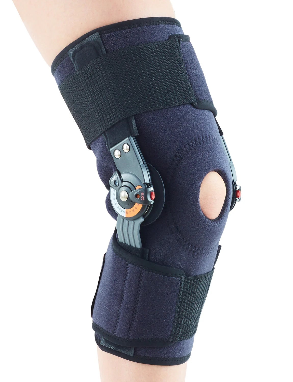
- Hinged Knee Brace
- Indications: Grade I, II, III
- Goal is to prevent further valgus injury
- Weight bearing as tolerated
- Advance as patient is pain free, able to walk without an antalgic gait
- NSAIDS
Operative management of MCL Tear
- Indications
- Grade 3 with other ligament injuries
- Grade 3 injuries at the tibial insertion
- Chronic MCL tears
- Technique
- Ligament repair
- Ligament reconstruction
Rehab and Return to Play
Rehabilitation
- General
- Early rehabilitation
- Range of motion, however prolonged immobilization can lead to weaker ligament healing, worse outcomes in animal models[20]
- Progressive strengthening
- Physical Therapy (early)
- Quad sets, straight leg raises, hip adduction
- Cycling
- Progressive resistance training
3 Phases of Therapy Goals
- Phase 1
- Protect the injury in hinged knee brace, rest
- Treat inflammation via swelling, ice, etc
- Other modalities: electrical simulation, ultrasound, other compression
- Work on knee motion, strength of surrounding muscles, normalize gait
- Phase 2
- Increasing strength
- Initiate regional therapy including core, hip abductors, external rotators, biomechanics
- Start aerobic activity, e.g. jogging in a straight line
- Phase 3
- Functional progression (running, agility, plyometrics, sports specific movements)
Return to Play/Work
- General rule for isolated MCL injuries
- Grade 1: 1-2 weeks
- Grade 2: 2-6 weeks
- Grade 3: 6-8 weeks
Prognosis and Complications
Prognosis
- Outcomes for grade I, II
- Typically good to excellent
- These athletes can return to previous level of activity
- Outcomes for grade III managed nonoperatively
- Some studies show good results, others show chronic laxity and early arthritis
- MCL injuries at tibial insertion tend to due worse than those of femoral origin
- Derscheid et al study of high school football players with nonoperative management of grade 1, 2 tears[21]
- Grade 1 tears returned to sport an average of 10.6 days post-injury
- Grade 2 tears returned an average of 19.5 days post-injury
Complications
- Instability/ Laxity
- More common after grade 2, 3 tears
- Persistent pain
- Rarely leads to Complex Regional Pain Syndrome
- Recurrence of injury
- In a group of patients with isolated grade 3 MCL injuries, the recurrence of MCL injury was 23%[22]
- Saphenous Nerve Injury
- Loss of range of motion
- Pellegrini Stieda Syndrome
See Also
Internal
External
- Sports Medicine Review Knee Pain: https://www.sportsmedreview.com/by-joint/knee/
References
- ↑ Brantigan OC, Voshell AF: The tibial collateral ligament: Its function, its bursae, and its relation to the medial meniscus. J Bone Joint Surg 25:121-131, 1943
- ↑ Kerlan, RK, Glousman, RE: Tibial collateral ligament bursitis. Am J Sports Med 1988;16:344–346.
- ↑ Majewski M., Susanne H., Klaus S. Epidemiology of athletic knee injuries: a 10-year study. Knee. 2006;13(3):184–188.
- ↑ Kim, Christopher, Patrick M. Chasse, and Dean C. Taylor. "Return to play after medial collateral ligament injury." Clinics in sports medicine 35.4 (2016): 679-696.
- ↑ Encinas-Ullán, Carlos A., and E. Carlos Rodríguez-Merchán. "Isolated medial collateral ligament tears: an update on management." EFORT open reviews 3.7 (2018): 398.
- ↑ Kramer, Dennis E., et al. "Collateral ligament knee injuries in pediatric and adolescent athletes." Journal of Pediatric Orthopaedics 40.2 (2020): 71-77.
- ↑ Jacob, George, et al. "Percutaneous arthroscopic assisted knee medial collateral ligament repair." Arthroscopy Techniques 9.10 (2020): e1511-e1517.
- ↑ Memarzadeh, Arman, and Joel TK Melton. "Medial collateral ligament of the knee: Anatomy, management and surgical techniques for reconstruction." Orthopaedics and Trauma 33.2 (2019): 91-99.
- ↑ Craft, Jason A., and Peter R. Kurzweil. "Physical examination and imaging of medial collateral ligament and posteromedial corner of the knee." Sports Medicine and Arthroscopy Review 23.2 (2015): e1-e6.
- ↑ Glousman, R. K. (1988). Tibial Collateral Ligament Bursitis. The American Journal of Sports Medicine, 344-346.
- ↑ De Maeseneer M, Shahabpour M, Van Roy F, Goossens A, De Ridder F, Clarijs J, Osteaux M. MR imaging of the medial collateral ligament bursa: findings in patients and anatomic data derived from cadavers. (2001) AJR. American journal of roentgenology. 177 (4): 911-7.
- ↑ Hakan Nur, A. A. (2018). Medial collateral ligament bursitis in a patient with knee osteoarthritis. Journal of Back and Musculoskeletal Rehabilitation, 589-591.
- ↑ Encinas-Ullán, Carlos A., and E. Carlos Rodríguez-Merchán. "Isolated medial collateral ligament tears: an update on management." EFORT open reviews 3.7 (2018): 398.
- ↑ Case courtesy of Frank Gaillard, Radiopaedia.org, rID: 5650
- ↑ Ghosh, N., et al. "Comparing point-of-care-ultrasound (POCUS) to MRI for the diagnosis of medial compartment knee injuries." Journal of medical ultrasound 25.3 (2017): 167-172.
- ↑ Phisitkul P., James S.L., Wolf B.R., Amendola A. MCL injuries of the knee: current concepts review. Iowa Orthop J. 2006;26:77–90.
- ↑ De Maeseneer, M, Shahabpour, M, Van Roy, F: MR imaging of the medial collateral ligament bursa: findings in patients and anatomic data derived from cadavers. Am J Roentgenol 2001;177:911–917.
- ↑ Vincenzo Ricci, L. O. (2019). Ultrasound‐Guided Treatment of Extrusive Medial Meniscopathy: A 3‐Step Protocol. Journal of Ultrasound in Medicine.
- ↑ Jean Jose, E. S. (2011). Sonographically Guided Therapeutic Injection for Primary Medial (Tibial) Collateral Bursitis. The Journal of Clinical Ultrasound, 257-261.
- ↑ Creighton R.A., Spang J.T., Dahners L.E. Basic science of ligament healing. Sports Med Arthrosc Rev. 2005;13(3):145–150.
- ↑ Derscheid G.L., Garrick J.G. Medial collateral ligament injuries in football: nonoperative management of grade I and grade II sprains. Am J Sports Med. 1981;9:365–368.
- ↑ Reider B., Sathy M.R., Talkington J., Blyznak N., Kollias S. Treatment of isolated medial collateral ligament injuries in athletes with early functional rehabilitation. Am J Sports Med. 1994;22(4):470–477.
Created by:
John Kiel on 7 July 2019 05:43:50
Authors:
Last edited:
27 March 2025 17:53:27
Categories: