LCL Injury
Other Names
- LCL Tear
- Lateral Collateral Ligament Tear
- Lateral Collateral Ligament Injury
- LCL Sprain
- Fibular ligament injury
Background
- This page refers to injuries to the Lateral Collateral Ligament
History
- Needs to be updated
Epidemiology
- Second least frequent of all ligamentous knee injuries at 7.9% (behind PCL)[1]
- Isolated knee injuries represent less than 2% of knee injuries[2]
Introduction

- Rarely occurs in isolation, most commonly occurs with Posterolateral Corner Injury
Etiology
- Contact sports
- Commonly medial blow to knee while in full extension
- Noncontact[4]
- Varus bending
- Hyperextension
- Motor vehicle crashes
Anatomy
- Lateral Collateral Ligament
- Originates from lateral femoral condyle and inserts onto the fibular head
- Primary varus stabilizer of the knee[5]
- Secondary restraint to external rotation, posterior displacement of Tibia
- Posterolateral Corner
- Formed by Lateral Collateral Ligament, popliteus muscle-tendon unit, popliteofibular ligament (PFL)
- Frequently co-injured with LCL injuries
Associated Conditions
- Meniscus Injury
- Osteochondral Defect
- Multiligament Knee Injury
- ACL Injury
- PCL Injury
- MCL Injury
- Posterolateral Corner Injury
- Bone contusion, less commonly fracture
- Knee Dislocation
Risk Factors
- Female gender
- Sports that require high velocity pivoting, jumping
- Sports[6]
- Tennis
- Gymnastics
- Wrestling
- Orthopedic history
- History of knee, ankle or hip injury[7]
Differential Diagnosis
- Fractures
- Dislocations & Subluxations
- Patellar Dislocation (and subluxation)
- Knee Dislocation
- Proximal Tibiofibular Joint Dislocation
- Muscle and Tendon Injuries
- Ligament Pathology
- Arthropathies
- Bursopathies
- Patellofemoral Pain Syndrome (PFPS)/ Anterior Knee Pain)
- Neuropathies
- Other
- Bakers Cyst (Popliteal Cyst)
- Patellar Contusion
- Pellegrini Stieda Syndrome
- Parameniscal Cyst
- Pediatric Considerations
- Patellar Apophysitis (Sinding-Larsen-Johnansson Disease)
- Patellar Pole Avulsion Fracture
- Tibial Tubercle Avulsion Fracture
- Tibial Tuberosity Apophysitis (Osgood Schalatters Disease)
- Proximal Tibial Metaphyseal Fracture
- Proximal Tibial Physeal Injury
Clinical Features

- History
- Generally patients will report an acute, traumatic event
- Sudden onset lateral knee pain, swelling and bruising
- Instability with knee in extension
- Trouble with stairs, cutting or pivoting activities
- Rarely weakness, parasthesia or foot drop (increase risk in PLC injuries)
- Sensation of walking "bow legged" due to increase laxity
- Physical Exam: Physical Exam Knee
- Tenderness to palpation of the distal lateral femur and/or fibular head
- Ecchymosis, swelling and warmth may be present
- Effusion is typically absent in isolated injuries (due to extra-articular nature of LCL)
- Special Tests
- Varus Stress Test (Knee): one hand on lateral knee, other on leg applying varus stress at 0° and 30°
- Posterolateral corner tests
- External Rotation Recurvatum Test: Patient supine, downward force on suprapatellar region while externally rotating tibia
- Posterolateral Drawer Test: Prone, knee flexed to 90°, externally rotated 15°
- Reverse Pivot Shift Test: Prone, knee 70° of flexion and the foot is rotated externally and slowly brought to 20-30°
- Dial Test: Prone, knees flexed to 30° with external rotation, then retested at 90°
- Important to perform thorough structural knee exam on MCL, ACL, PCL, posterolateral corner, etc


Evaluation
Radiographs
- Standard Radiographs Knee
- Routine screening
- Typically normal
- May demonstrate
- Fibular head fractures/avulsions (arcuate sign)
- Tibial spine avulsions
- Lateral tibial plateau (i.e., Segond fracture)
- Alternative views
- Varus stress view
- Kneeling posterior stress radiographs
- Measure shortest distance between most distal aspect of lateral femoral condyle, associated tibial plateu
MRI
- Gold standard for evaluating LCL
- Best seen in axial, coronal views
- Accuracy (need citation)
- Sensitivity: 90%
- Specificity: 90%
- Primary findings
- Majority of tears off fibular insertion
- Medial compartment bone bruising (T2-weighted)
- Varying amounts of peri-ligamentous edema, intra-substance signal
- Discontinuity of LCL fibers in higher grade tears
US
- Acute findings
- Edema
- Loss of fiber continuity
- Dynamic laxity
- Chronic findings
- Thickened
- Hypoechoic
Classification
LCL Tear Classification
- Grade 1 (Mild)
- Localized lateral knee tenderness
- No instability or mechanical symptoms are present.
- 0 to 4 mm of laxity
- Grade 2 (Partial Tear)
- Severe localized lateral and posterolateral knee pain, swelling.
- 5 to 10 mm of laxity with a fixed endpoint
- Grade 3 (Complete Tear)
- Pain and swelling vary in patients
- Usually associated with PLC and other related injuries
- >10mm of laxity with no firm end point
LCL Tear MRI Classification
- Grade 1
- Subcutaneous fluid surrounding the midsubstance of the ligament at one or both insertions
- Grade 2
- Partial tearing of ligament fibers at either the midsubstance or one of the insertions
- Increased edema
- Grade 3
- Complete tearing of ligament fibers at either the midsubstance or one of the insertions
- Increased edema
Management
Nonoperative
- Indications[11]
- Grade I, II
- Stable varus stress test at 0°
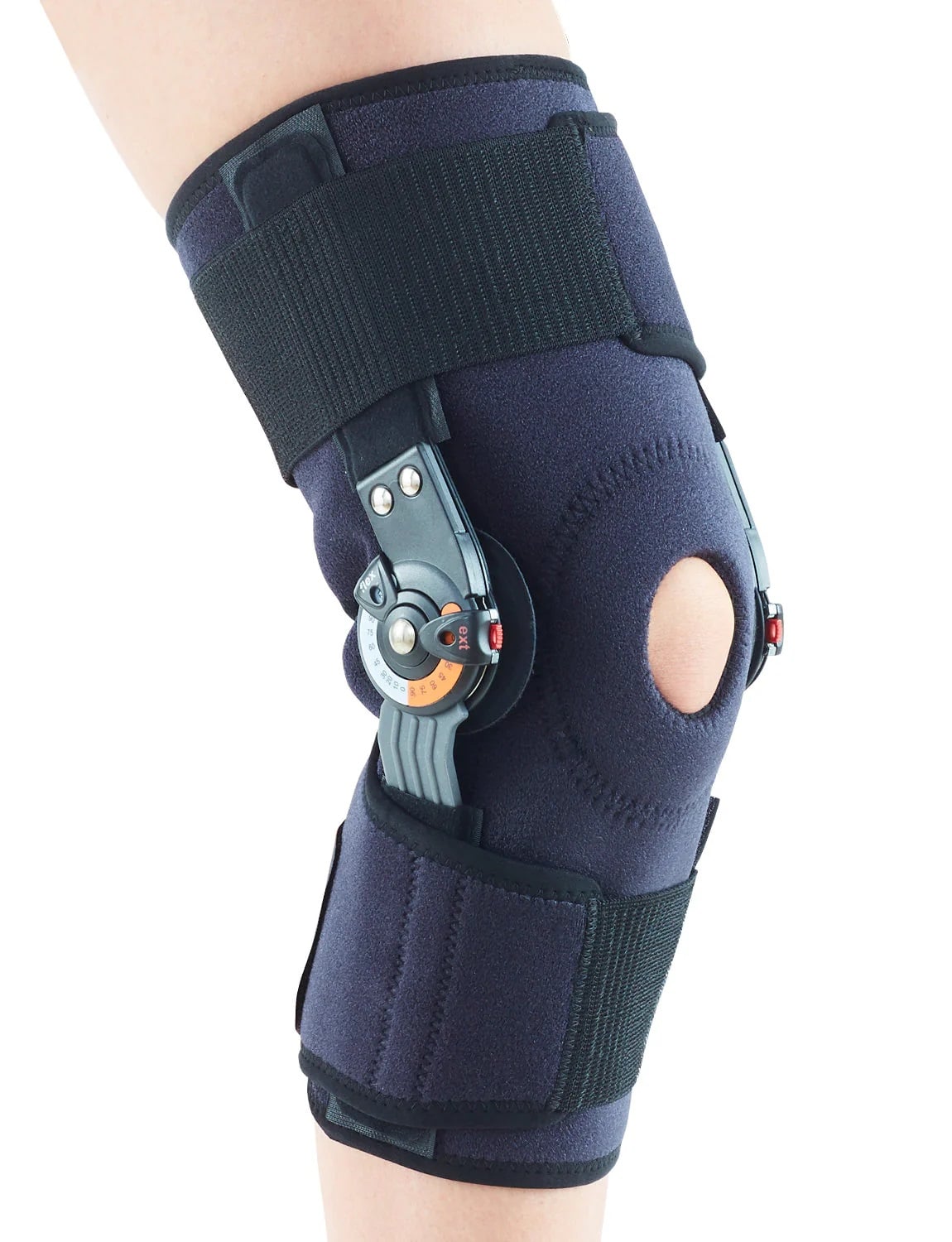
- Hinged Knee Brace
- Should restrict varus motion
- Discontinue after pain, sensation of instability resolve
- Early Mobilization
- Physical Therapy
- Not always necessary in isolated injuries
- May be needed in cases of prolonged immobilization
Operative
- Indications
- Grade III injuries (controversial)
- Co-occuring injuries such as posterolateral corner, multiligament injuries
- Technique
- Repair
- Reconstruction
Rehab and Return to Play
Rehabilitation
- General
- Limited immobilization with progression range of motion
- functional rehabilitation
- Emphasis on quadriceps, hamstring strengthening
Return to Play/ Work
- Grade I and II
- Can often return to sport in 6-8 weeks
Complications and Prognosis
Prognosis
- Bushnell at al looked at NFL players with isolated grade III LCL injuries[2]
- Nonsurgically managed injuries were as likely as those with surgically managed injuries to return to professional play
- They did so more quickly
- Krukhuag et al found surgically managed isolated grade III LCL injuries did better than nonsurgical counterparts[12]
- Kannus found nonoperative management of isolated grade III injuries did not do as well as surgically managed patients[11]
- Findings: severe or gross lateral laxity, insufficiency of the ACL, muscle weakness, and posttraumatic osteoarthritis
- Studies show that the LCL does not heal as well as the MCL (need citation)
Complications
- Common Peroneal Nerve Injury
- Occurs in up to 44% of LCL/PLC combined injuries[4]
- Persistent varus or hyperextension laxity
- Stiffness
- Tends to occur following prolonged immobilization with nonoperative management
- Physeal arrest
- Can be seen in skeletally immature patient with errant lateral condylar LCL fixation
See Also
Internal
External
- Sports Medicine Review Knee Pain: https://www.sportsmedreview.com/by-joint/knee/
References
- ↑ Swenson DM, Collins CL, Best TM, Flanigan DC, Fields SK, Comstock RD. Epidemiology of knee injuries among U.S. high school athletes, 2005/2006-2010/2011. Med Sci Sports Exerc. 2013 Mar;45(3):462-9.
- ↑ 2.0 2.1 Bushnell BD, Bitting SS, Crain JM, Boublik M, Schlegel TF: Treatment of magnetic resonance imaging-documented isolated grade III lateral collateral ligament injuries in National Football League athletes. Am J Sports Med 2010;38(1):86-91.19966106
- ↑ LaPrade RF, Spiridonov SI, Coobs BR, Ruckert PR, Griffith CJ (2010) Fibular collateral ligament anatomical reconstructions: a prospective outcomes study. Am J Sports Med 38(10):2005–2011
- ↑ 4.0 4.1 Chahla, Jorge, et al. "Posterolateral corner of the knee: current concepts." Archives of Bone and Joint Surgery 4.2 (2016): 97.
- ↑ Wilson WT, Deakin AH, Payne AP, Picard F, Wearing SC: Comparative analysis of the structural properties of the collateral ligaments of the human knee. J Orthop Sports Phys Ther 2012;42(4):345-351.22030378
- ↑ Grawe, Brian, et al. "Lateral collateral ligament injury about the knee: anatomy, evaluation, and management." JAAOS-Journal of the American Academy of Orthopaedic Surgeons 26.6 (2018): e120-e127.
- ↑ Hill OT, Bulathsinhala L, Scofield DE, Haley TF, Bernasek TL. Risk factors for soft tissue knee injuries in active duty U.S. Army soldiers, 2000-2005. Mil Med. 2013 Jun;178(6):676-82
- ↑ Jain, Rajat K. "Lateral collateral ligament injury." Common Pediatric Knee Injuries: Best Practices in Evaluation and Management (2021): 225-232.
- ↑ Case courtesy of Haytham Mohamed Assayed Bedier, Radiopaedia.org, rID: 56857
- ↑ Case courtesy of Henry Knipe, Radiopaedia.org, rID: 62164
- ↑ 11.0 11.1 Kannus, Pekka. "Nonoperative treatment of grade II and III sprains of the lateral ligament compartment of the knee." The American Journal of Sports Medicine 17.1 (1989): 83-88.
- ↑ Krukhaug Y, Mølster A, Rodt A, Strand T. Lateral ligament injuries of the knee. Knee Surg Sports Traumatol Arthrosc. 1998;6(1):21-5.
Created by:
John Kiel on 7 July 2019 05:43:53
Authors:
Last edited:
5 August 2024 17:35:01
Categories: