Patellofemoral Joint
Description
-
![Patellofemoral joint (PFJ) arthritis. A. Skyline X-Ray showing PFJ osteoarthritis. B. Skyline X-ray after PFJ replacement. C. Lateral X-ray showing PFJ osteoarthritis. D. Postoperative lateral X-ray after PFJ arthroplasty. E. Anteroposterior X-ray after PFJ arthroplasty[1]](/w/images/thumb/5/50/Patellofemoral_joint_replacement_xray.jpeg/120px-Patellofemoral_joint_replacement_xray.jpeg) Patellofemoral joint (PFJ) arthritis. A. Skyline X-Ray showing PFJ osteoarthritis. B. Skyline X-ray after PFJ replacement. C. Lateral X-ray showing PFJ osteoarthritis. D. Postoperative lateral X-ray after PFJ arthroplasty. E. Anteroposterior X-ray after PFJ arthroplasty[1]
Patellofemoral joint (PFJ) arthritis. A. Skyline X-Ray showing PFJ osteoarthritis. B. Skyline X-ray after PFJ replacement. C. Lateral X-ray showing PFJ osteoarthritis. D. Postoperative lateral X-ray after PFJ arthroplasty. E. Anteroposterior X-ray after PFJ arthroplasty[1] -
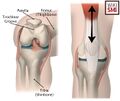
![Six degrees of freedom movement of the patella relative to the femorotibial joint[2]](/w/images/thumb/8/83/Patella_movement.jpeg/120px-Patella_movement.jpeg) Six degrees of freedom movement of the patella relative to the femorotibial joint[2]
Six degrees of freedom movement of the patella relative to the femorotibial joint[2] -
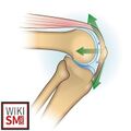
![Schematic indices of patellofemoral joint anatomy[3]](/w/images/thumb/5/51/Patellofemoral_joint_anatomy.jpeg/120px-Patellofemoral_joint_anatomy.jpeg) Schematic indices of patellofemoral joint anatomy[3]
Schematic indices of patellofemoral joint anatomy[3] -
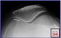 Axial radiograph illustrating medial patellofemoral joint arthritis
 -
 Patellofemoral joint anatomy
 -
 Patellofemoral joint anatomy
![Patellofemoral joint (PFJ) arthritis. A. Skyline X-Ray showing PFJ osteoarthritis. B. Skyline X-ray after PFJ replacement. C. Lateral X-ray showing PFJ osteoarthritis. D. Postoperative lateral X-ray after PFJ arthroplasty. E. Anteroposterior X-ray after PFJ arthroplasty[1]](/File:Patellofemoral_joint_replacement_xray.jpeg)
![Six degrees of freedom movement of the patella relative to the femorotibial joint[2]](/File:Patella_movement.jpeg)
![Schematic indices of patellofemoral joint anatomy[3]](/File:Patellofemoral_joint_anatomy.jpeg)


Name
- Patellofemoral Joint
- Kneecap Joint
- Patellar joint
- Patellar–femoral articulation
- Femoro–patellar joint
- Patellofemoral articulation
- Kneecap joint (informal)
- Anterior knee joint compartment (anatomical description)
- Patellofemoral compartment of the knee
General
- Biomechanically complex
- Synovial joint between the condyles of the Femur, articular surface of Patella
- Contiguous and part of the the Knee Joint
- Posterior patella: medial, lateral and odd facets articulate; lateral is largest and steepest
- Femur: medial and lateral condyles articulate
- Articular surface covered with Articular Cartilage
- Joint capsule is contiguous with the knee joint
Static stabilizers
Dynamic stabilizers
- Quadriceps Femoris proximally via the Quadriceps Tendon
- Patellar Tendon on the inferior pole attaches to the tibial tubercle
- Vastus Medialis: medial restraint to lateral translation
- Vastus Lateralis: lateral restraint to medial translation
Actions
- Knee extension
- Patellar slides along femoral condyles
- Increases lever arm of the extensor mechanism
- Transmits tensile forces generated by the quadriceps to the patellar tendon
Vascular Supply
- Branches of the Popliteal Artery including
- Superior, inferior medial, lateral geniculate arteries
- Branches of the Femoral Artery including
- Descending geniculate branches
Innervation
- Branches of
Clinical Significance
Atraumatic
- Patellofemoral Pain Syndrome
- Chondromalacia Patellae
- Patellofemoral Joint Arthritis
- Patellar Instability
Traumatic
- Patella Dislocation
- Patella Fracture
- Patella Tendon Rupture
- Quadriceps Tendon Rupture
- Osteochondral Defect
See Also
References
- ↑ Ahmad, Sufian S., et al. "Arthroplasty–current strategies for the management of knee osteoarthritis." Swiss medical weekly 145.0708 (2015): w14096-w14096.
- ↑ Xue, Zhe, et al. "Development of an innovative measurement method for patellar tracking disorder." Aging (Albany NY) 13.1 (2020): 516.
- ↑ Leiprecht, J., et al. "Weight-bearing MRI with a knee flexion angle of 20: a study on additional MRI investigation modalities to support a more accurate understanding of patellofemoral instability." BMC Musculoskeletal Disorders 22.1 (2021): 902.<
- ↑ Image courtesy of kenhub.com
- ↑ Masroori, Zahra, et al. "Patellar Non-Traumatic Pathologies: A Pictorial Review of Radiologic Findings." Diagnostics 14.24 (2024): 2828.